Cell Proliferation and Viability Assay
Enhancing Cellular Viability Analysis for Drug Discovery
At Sygnature Discovery, we have harnessed a diverse array of techniques to help you uncover biologically active compounds that illuminate the effects of target inactivation on cell proliferation, activation, apoptosis, or cytotoxic cell death.
Exploring Cell Proliferation and Viability Analysis
Cell proliferation and viability assays serve as robust tools, playing a pivotal role in the discovery of novel therapeutic agents. Most in vitro proliferation assays not only determine the number of viable cells but also encompass methods for quantifying cell nuclei, assessing metabolic activity, gauging cell redox potential, and evaluating mitochondrial integrity.
These versatile proliferation assays can be seamlessly integrated with cytotoxicity or apoptosis assays, enabling real-time monitoring of cellular responses to compound treatments. Compound-induced cell apoptosis can be quantified through indicators like an increase in cell membrane permeability, the loss of membrane asymmetry, or the activation of caspases.
Our automated liquid handling systems facilitate high-throughput data acquisition, expediting compound validation and aiding selection for subsequent screening cycles within the Design-Make-Test-Analyse framework. These methodologies for assessing cell proliferation and viability find application across both Phenotypic and target-driven drug discovery projects.
Noteworthy applications include:
- Peripheral blood mononuclear cells (PBMCs) obtained from our Blood Donor Panel
- Primary cell populations or subpopulations (T cells, B cells, Natural Killer cells, macrophages)
Diverse Techniques for Cellular Proliferation, Viability, and Apoptosis Evaluation
Our arsenal of techniques encompasses a range of methods for quantifying cell proliferation, viability, and apoptosis, including:
- Detection of cell’s metabolic activity
- Resazurin reduction
- ATP luciferase coupled reaction
- Quantification of cell nuclei or cell confluency
- Fluorescent labelling of DNA/cell nuclei
- Acquisition of phase contrast images of cells using IncuCyte® ZOOM
- Detection of actively dividing cells
- BrdU incorporation into newly synthesized DNA
- Cell apoptosis/cytotoxicity
- Caspase 3/7 activation during cell apoptosis
- Increase in cell membrane permeability
- Loss in cell membrane asymmetry by detection of exposed phosphatidylserine on cell surface
Illustrating the Significance of Cellular Viability and Proliferation Analysis
Examples of in vitro assays to measure a cell proliferation assay, viability and apoptosis.
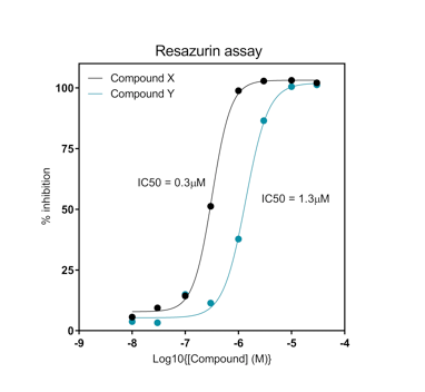
Figure A: Resazurin assay using cells treated with two compounds. Compounds display different potency on proliferation of cells.
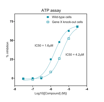
Figure B: ATP Luciferase coupled assay. Compounds display different potency on proliferation of wild-type versus knock-out cells.
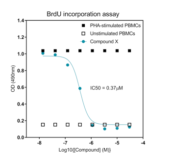
Figure C: BrdU incorporation assay with PMBCs that were left unstimulated or stimulated with PHA in the presence of selected compound. The compound reduces activation and proliferation of T-cells responding to stimulus.
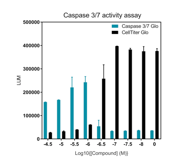
Figure D: Caspase 3/7 Glo® activities in cells treated with a compound. Data expressed in comparison to CellTiter Glo® to demonstrate correlation between a reduction in cell viability with an increase in caspase 3/7 activation.
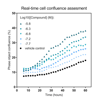
Figure E: Real-time cell confluency assessment using the IncuCyte® ZOOM instrument. Data display a time-dependent increase in cell confluency in response to stimulation.
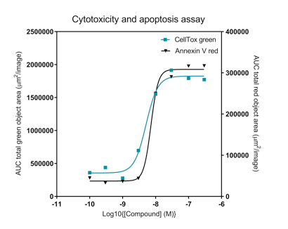
Figure F: Detection of cytotoxicity and apoptosis in cells treated with a compound. Data shows dose-dependent increase in CellTox Green® intracellular accumulation and Annexin V binding in response to compound treatment.
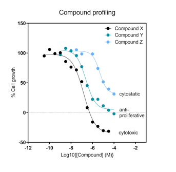
Figure G: Determination of compound effect on cell growth. The graph shows a compound that is cytotoxic (X), anti-proliferative (Y) or cytostatic (Z).
In summary, our comprehensive suite of methodologies empowers refined analyses of cell viability and proliferation, underpinning advances in drug discovery and furthering our understanding of cellular responses to therapeutic interventions.
