

Featured Resources
-
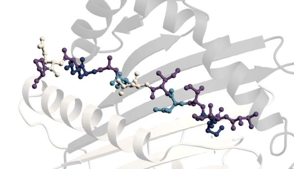
STORM Therapeutics: From Lead to Pre-Candidate Nomination in 18 Months

STORM Therapeutics: From Lead to Pre-Candidate Nomination in 18 Months
Case study
📣From lead to pre-candidate nomination in 18 months. Explore the STORM Therapeutics case study


Discover precise insights into brain neurochemistry with Sygnature Discovery's in vivo microdialysis and cOFM services. With over 20 years of expertise, we design bespoke studies that reveal how compounds modulate neurotransmitter systems in health and disease. Using UHPLC/HPLC with electrochemical detection or mass spectrometry, we deliver robust PK/PD data to support confident CNS decision making.


July 30, 2015

Inobrodib, an exciting, first-in-class oral anti-cancer drug in clinical development by CellCentric, was collaboratively designed, synthesised and supported on its pre-clinical journey by an integrated project team at Sygnature Discovery. Inobrodib is now showing promising results in Phase I and II trials for multiple myeloma and other cancer types.

At Sygnature Discovery, we deliver world-leading drug discovery solutions to accelerate your compound from idea to clinic.

Our leadership team brings diverse experience and insight, driving collaboration and innovation across drug discovery.

Explore careers at Sygnature Discovery and join a global team committed to science, collaboration, and integrity.
Inobrodib, an exciting, first-in-class oral anti-cancer drug in clinical development by CellCentric, was collaboratively designed, synthesised and supported on its pre-clinical journey by an integrated project team at Sygnature Discovery. Inobrodib is now showing promising results in Phase I and II trials for multiple myeloma and other cancer types.
At Sygnature Discovery, we deliver world-leading drug discovery solutions to accelerate your compound from idea to clinic.
Our leadership team brings diverse experience and insight, driving collaboration and innovation across drug discovery.
Explore careers at Sygnature Discovery and join a global team committed to science, collaboration, and integrity.
Bioscience | cell-based assays
Cell-based assays drive smarter decisions in drug discovery from early validation to late-stage studies.

Our process
Developing robust and relevant in vitro and ex vivo cell assay solutions is essential for making informed decisions at the right time. At Sygnature Discovery, our team of expert cell biologists design tailored assays to fully characterize and validate your targets, helping you advance compounds from hit validation through candidate selection.

We offer a comprehensive suite of cell-based assays. These solutions enable precise profiling and characterization across diverse biological systems.

Comprehensive ion channel assays and bespoke cell lines deliver high-fidelity screening and safety insights, accelerating hit-to-lead and optimization.

End-to-end GPCR expertise combines advanced assays and structural biology to accelerate candidate selection and deliver confident, data-driven decisions.

Advanced transporter assays and electrophysiology deliver robust, high-throughput data for hit identification, validation, and lead optimization in drug discovery.

Robust binding, signaling and functional assays enable precise evaluation of receptor affinity and mechanism of action, supporting drug design and optimization.

Custom reporter gene assays integrated into stable cell lines enable quantitative measurement of receptor pathway activation and signalling mechanisms.

Multi-parametric imaging/screening of 3D cell cultures enables quantitative evaluation of compound-dependent effects across clinically relevant parameters.

Phenotypic screens using immortalised and primary cell models deliver contextually relevant, data-rich insight into novel targets and chemical matter.

Sensitive gene and protein expression assays provide quantitative, target-specific data to characterize cellular responses and inform mechanism of action.
Choosing the right assay is critical to your program’s success. Early in discovery, recombinant overexpression cell assays are often ideal, they’re high-throughput, easy to interpret, and optimized for identifying compounds that interact with your target. These systems can include related family members for selectivity studies or species homologues for cross-reactivity assessments.
As programs progress, confirming target engagement in physiologically relevant cells become essential. That’s why we provide assays using human iPSC, primary cells and ex vivo tissues. Our scientists design complex phenotypic assays in 2D and 3D formats, characterizing signalling and expression patterns to predict functional outcomes. Medium- to high-throughput assays allow rapid profiling of novel compounds, gene expression analysis, and biomarker identification.
Right Assay, Right Time
Every program is unique. We collaborate with you to develop and optimize custom assays to meet your specific needs. Our capabilities span:
qPCR, qRT-PCR, high content imaging, Jess Simple Western, Flow Cytometry, western blotting, protein degradation
Whole cell fluorescent ligand binding, receptor internalization
e.g., Ca2+ mobilisation, cAMP accumulation, phospho-ERK
Cell viability/proliferation, apoptosis, cell morphology, migration/invasion, differentiation, cell binding and internalisation
Selecting the right cell system is the first step. We offer:
Overexpressed targets for sensitive detection.
Cancer lines, human primary cells, iPSC-derived cells
Our team works with you to design assays that monitor ligand binding, intracellular signalling, and functional outcomes. Combining data from multiple assay types accelerates progression from hit-to-lead through preclinical proof-of-concept.
Resource hub


We bring together a dynamic blend of new talent and experiences pharmaceutical industry experts from around the world, delivering excellence across each stage of discovery.

Custom stable cell lines with robust expression and assay reproducibility, streamlined design and validation for confident drug discovery workflows.

Integrated hit identification using HTS, fragment screening, and advanced automation delivering high-quality starting points for successful drug discovery.

High-throughput biophysical solutions for binding, kinetics, and target engagement combining advanced technologies with deep expertise to guide drug discovery.
Custom assay solutions built around your biology delivering reliable, decision-ready data to advance hit finding and compound profiling with confidence.

Access to primary cells and tissues for physiologically relevant assays enabling confident, clinically predictive decisions earlier in drug discovery.
Custom biochemical assays built around your target biology providing robust, high-quality data to guide hit identification and mechanism-of-action studies.
Sygnature Discovery prides itself on offering a unique, high-quality, distinctive and tailored integrated drug discovery service to our clients.
Sygnature Vendor onsite cell bank offer > 100 cell lines spanning lung, breast, colorectal, ovarian, prostate cancer and more. Our cell lines span human, mouse, rat and monkey cell lines. We can generate recombinant cell lines in house and have access to human primary cells or iPSC-derived cell lines from external vendors. Fresh whole blood is obtained from our internal blood donor bank or through NHS Blood and Transplant (NHSBT).
If we don’t currently have access to the most appropriate cell-line for your research, Sygnature will work with you to arrange access to the relevant licenses.
Our range of plate readers, liquid handling systems and imagers can accommodate all standard formats from slides through to 96, 384 or 1536 well plates.
Combined with choice of automation solutions, we can offer high throughput screening approaches for a variety of cell-based assays.
Sygnature Discovery offers a comprehensive suite of technologies to support a broad spectrum of quantitative endpoints in cell-based assays.
Our platforms enable precise measurement of cellular protein expression and localization through Jess Automated Western Blotting and High Content Imaging using confocal and widefield immunofluorescence systems. For secreted proteins and biomarkers, we provide multiplex quantification via Luminex, alongside ELISA and Lumit® based detection methods.
We bring deep expertise in homogeneous assay formats, including AlphaLISA, HTRF, HiBiT, and Calcium Flux, ensuring sensitive and scalable readouts. Gene expression analysis is streamlined with our high-throughput, 384 well automated qPCR workflows.
In addition, Sygnature offers robust solutions for assessing cell viability and proliferation in both 2D and 3D models – empowering your research with reliable, reproducible data across diverse biological contexts.
Peak Proteins, NuChem Sciences, and SB Drug Discovery have now fully integrated with Sygnature Discovery.


