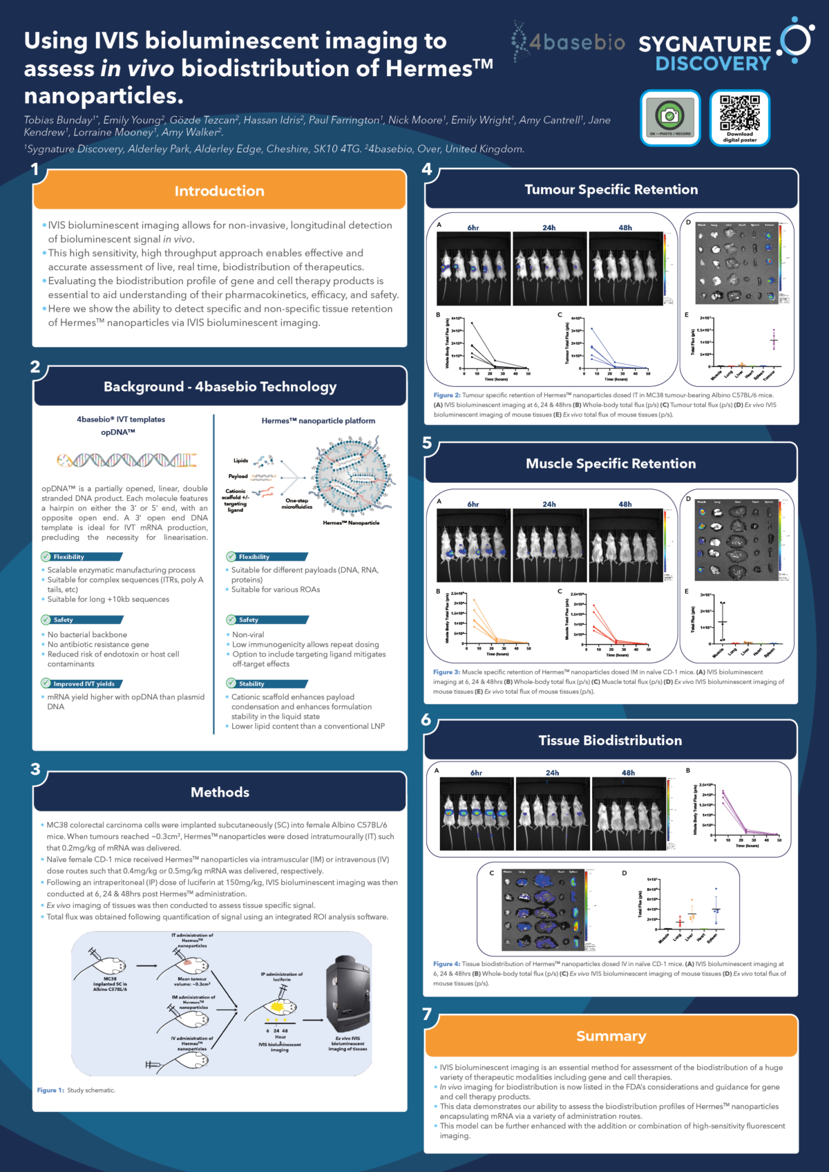
Using IVIS bioluminescent imaging to assess in vivo biodistribution of HermesTM nanoparticles
IVIS bioluminescent imaging allows for non-invasive, longitudinal, detection of in vivo bioluminescent signal. This high sensitivity, high throughput approach enables effective and accurate assessment of live, real time, biodistribution of therapeutics. Evaluating the biodistribution profile of gene and cell therapy products is essential to aid understanding of their pharmacokinetics, efficacy, and safety. Moreover, methodologies for evaluating biodistribution profiles, including in vivo imaging, are now listed in the FDA’s considerations and guidance for gene and cell therapy products. Additionally, live imaging allows for the reduction of animal numbers, and can remove the need to periodically sample animal tissues for plate-based biodistribution assays.
4basebio designs, manufactures, and supplies application specific synthetic DNA or mRNA. Hermes™, their non-viral delivery system, encapsulates these payloads for vaccine and gene therapy applications. 4basebio’s scalable, fully enzymatic linear DNA synthesis process via Trueprime™ amplification technology enables production of synthetic DNA constructs devoid of bacterial backbone or an antibiotic resistance marker. The technology is size and sequence independent, enabling the incorporation of polyA tails >120 bp, and allows for large scale production of linear DNA with improved yields over traditional plasmid DNA fermentation processes. opDNA™ replaces plasmid DNA as a template during the in vitro transcription of mRNA, demonstrating higher mRNA yields and avoiding the need for an enzymatic linearisation step. Hermes™ employs both traditional lipids and a cationic ligand to drive payload encapsulation and stability in the liquid state.
Using IVIS bioluminescent imaging, we demonstrate the ability to assess the biodistribution profiles of HermesTM nanoparticles encapsulating mRNA which encodes firefly luciferase, following either intratumoural (IT), intramuscular (IM) or intravenous (IV) administration. For IT, albino C57BL/6 mice were implanted subcutaneously with MC38 cells on the left flank, and subsequently received 0.2mg/kg mRNA upon establishment of tumours. For IM and IV, non-tumour bearing CD-1 mice received 0.4mg/kg mRNA via the hindlimb muscle, or 0.5mg/kg mRNA via the tail vein, respectively. Imaging was carried out at 6, 24 and 48hrs post dose. Upon termination, organs were excised and ex vivo imaging conducted, to assess tissue specific bioluminescent signal. Total flux was obtained following quantification of bioluminescent signal using an integrated ROI analysis software.
In mice receiving an IT dose, localisation of bioluminescent signal only within the tumour was observed, indicating tumour specific retention of HermesTM nanoparticles. Similarly, in mice receiving an IM dose, signal localisation was observed in the hindlimb muscle only, again indicating retention within the specific tissue in which nanoparticles were administered. In mice receiving an IV dose, strong signal localisation was observed in the liver, lungs, and spleen. Moreover, specific biodistribution to non-hepatic tissues could be manipulated through modification of Hermes™ lipid and ligand molar ratios. In all animals, the ex vivo image correlated with the live images obtained. This highlights the accuracy and sensitivity of IVIS bioluminescent imaging and illustrates the ability to detect therapeutics targeting specific tissues.

