Exploring lysosome function in fibroblasts from Alzheimer’s and Parkinson’s disease patients: a platform for drug discovery
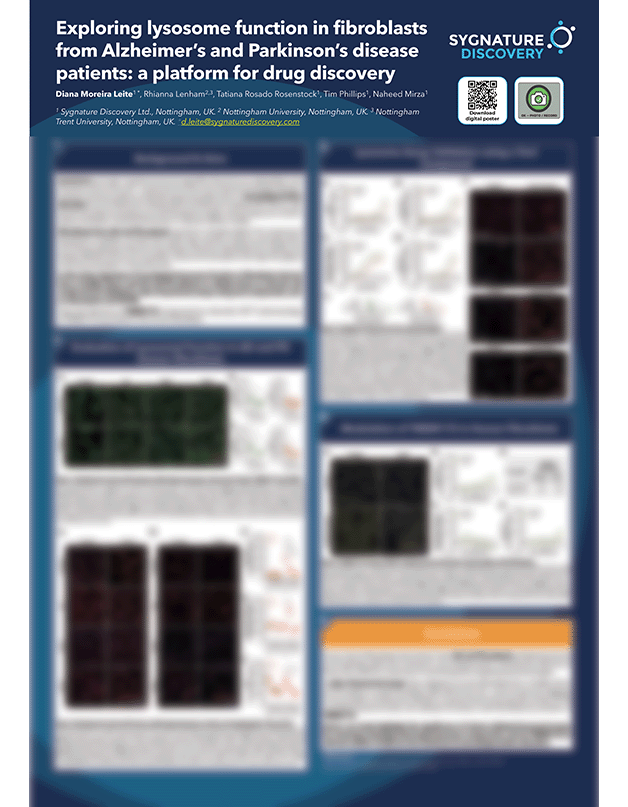
Lysosomes are acidic membrane-bound organelles that serve as a degradation and recycling centre of the cell, while also functioning as a signalling hub and participating in various biological processes, such as membrane repair, homeostasis, and immune response. Growing evidence implicates lysosomal dysfunction in neurodegenerative disorders, including Alzheimer’s (AD) and Parkinson’s disease (PD), and with lysosomal proteins emerging as novel therapeutic targets, robust assays are essential to assess modulation of lysosomal function in relevant disease cell models. Here, we initially evaluated human dermal fibroblasts derived from a healthy donor, and AD and PD patients to assess whether there are differences in aspects of lysosomal function. Assays to quantify lysosome numbers, including LAMP1 and LysoTrackerTM staining, pH (LysoSensor), enzymatic activity (cathepsin B) and autophagy initiation (p62 staining) were employed. Fibroblasts from AD and PD patients displayed reduced levels of lysosome numbers and altered autophagic flux when compared to the healthy donor, particularly, when under serum starvation. However, no significant differences were found in the lysosomal pH or cathepsin B activity. We used fibroblasts from a healthy donor and a PD patient to screen modulators of lysosomal activity, including chloroquine and DCPIB – an activator of the lysosomal membrane protein TMEM175, in a 384-well high-throughput format. Chloroquine, used as a tool compound, triggered an increase in LysoTrackerTM, lysosome de-acidification, and increased cathepsin B activity and p62 levels, reflecting altered lysosome function and inhibition of autophagy in both healthy and PD fibroblasts. With the TMEM175 activator DCPIB, an increase in LysoTrackerTM accumulation was observed in both healthy and PD fibroblasts (EC50 7 µM) along with de-acidification of lysosomes, reduced cathepsin enzyme B activity and autophagic flux. These data indicate that human dermal fibroblasts from patients present aspects of lysosomal and autophagic dysfunction reported in neurodegeneration. Further, our suite of assays can be used to probe lysosomal function and the effect of pharmacological modulators targeting different mechanisms within the lysosomes.

