

Featured Resources
-
STORM Therapeutics: From Lead to Pre-Candidate Nomination in 18 Months

STORM Therapeutics: From Lead to Pre-Candidate Nomination in 18 Months
Case study
📣From lead to pre-candidate nomination in 18 months. Explore the STORM Therapeutics case study


Discover precise insights into brain neurochemistry with Sygnature Discovery's in vivo microdialysis and cOFM services. With over 20 years of expertise, we design bespoke studies that reveal how compounds modulate neurotransmitter systems in health and disease. Using UHPLC/HPLC with electrochemical detection or mass spectrometry, we deliver robust PK/PD data to support confident CNS decision making.


July 30, 2015

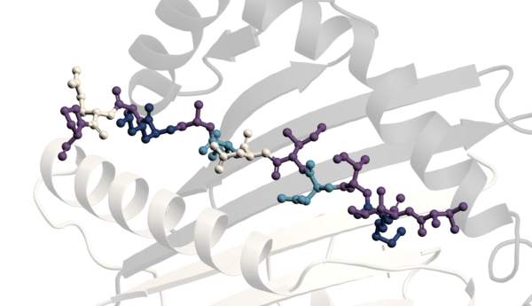
Inobrodib, an exciting, first-in-class oral anti-cancer drug in clinical development by CellCentric, was collaboratively designed, synthesised and supported on its pre-clinical journey by an integrated project team at Sygnature Discovery. Inobrodib is now showing promising results in Phase I and II trials for multiple myeloma and other cancer types.

At Sygnature Discovery, we deliver world-leading drug discovery solutions to accelerate your compound from idea to clinic.

Our leadership team brings diverse experience and insight, driving collaboration and innovation across drug discovery.

Explore careers at Sygnature Discovery and join a global team committed to science, collaboration, and integrity.
Inobrodib, an exciting, first-in-class oral anti-cancer drug in clinical development by CellCentric, was collaboratively designed, synthesised and supported on its pre-clinical journey by an integrated project team at Sygnature Discovery. Inobrodib is now showing promising results in Phase I and II trials for multiple myeloma and other cancer types.
At Sygnature Discovery, we deliver world-leading drug discovery solutions to accelerate your compound from idea to clinic.
Our leadership team brings diverse experience and insight, driving collaboration and innovation across drug discovery.
Explore careers at Sygnature Discovery and join a global team committed to science, collaboration, and integrity.
HTS is a ubiquitous and well-established approach for hit identification; it is often used in conjunction with other techniques, such as virtual and fragment-based drug discovery, for a target-based screening approach. Phenotypic screening, however, is used to identify new molecular target(s) or mechanisms and/or hits that modulate complex disease mechanisms in a desirable way.
The mechanism of a hit target interaction and the target’s identity are initially unknown post-phenotypic screening. This could be because the signalling cascade or disease process is poorly understood. The advantages of using this approach are that it delivers active molecules within a disease relevant cellular background, inclusive of solubility and cell permeability, providing candidates for further medicinal chemistry optimization. Phenotypic screening approaches are often aimed at delivering first-in-class compounds with disease-modifying modalities (Heilker, Lessel and Bischoff, 2019). Usually, developing an appropriate phenotypic assay is a compromise between achieving HTS assay practicality whilst maintaining a strong assay-disease linkage.
NLRP1 (aka NALP1) was the first described inflammasome sensor of the Nod-like Receptors (NLR) family, (Taabazuing, Griswold and Bachovchin, 2020). NLRP1 is an innate pattern recognition receptor; it senses molecules that pose a danger to the cell (i.e., bacterial toxins, stress factors and viral proteases). DPP-8/-9 (a dipeptidyl peptidase) holds NLRP1 in an auto-inhibitory state until stimulation/release, a process that can be artificially induced by Val-boroPro (VbP), a small-molecular inhibitor of DPP-8/-9 activity.
When active, NLRP1 facilitates inflammasome assembly, enabling activation of caspase-1, which, in turn, leads to the activation and release of IL-1ß, IL-18 and Gasdermin (which undergoes oligomerization and forms pores in the membrane), culminating in pyroptic cell death. There is considerable interest in the identification of inhibitors of excessive NLRP1 activity associated with a wide range of chronic inflammatory diseases, like IBD and asthma. Whilst NLRP1 activators are being explored as an immuno-oncology approach to treat cancer.
Several pathways can trigger NLRP1 activation that are unique to this class of inflammasome: –
We used a phenotypic screening approach for this target, considering the complex multimeric nature of NLPR1 and the technical difficulties in recapitulating an NLRP1 activation model using biochemical assay platforms. Binding type assays could potentially be used with purified NLRP1 or its domains (NACHT, FIIND, CARD, etc) but hits would not necessarily translate to functional inhibitors of NLRP1 inflammasome activation.
Here, we developed a High Content Screening assay where ASC, one of the components of the inflammasome, was labelled with GFP enabling facile visualization of intracellular inflammasome formation as a punctate ‘speck.’
This cell-based phenotypic assay provided an opportunity to evaluate NLRP1 activation (and inhibition) in a holistic cellular context, with the potential to identify modulators acting via multiple mechanisms. Often, when screening phenotypically, a disease relevant or patient derived cell line can be used to make the screen proximal to the disease state. In this screen, we used a cell line supplied by Invivogen. A549 is a lung carcinoma epithelial cell line, endogenously expressing proteins involved in the inflammasome signalling including ASC, caspase-1, and Gasdermin D/E.

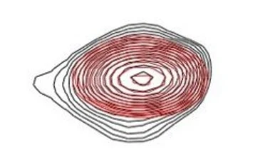
Figure 1 Schematic of inflammasome formation, including GFP-labelled ASC, used to visualize the inflammasome as a ‘speck.’

Figure 2 Representative images of A549 cells expressing ASC::GFP ± VbP induction. Cells were stained with Hoescht to visualize nuclei.

Figure 3 Graphs showing the effect of VbP on cell nuclei count, intracellular speck count and the calculated specks/cell ratio
We have established a fixed endpoint phenotypic cell assay to identify potential modulators of NLRP1-mediated inflammasome formation.
A 5000-compound subset of the Sygnature Discovery LeadFinder Diversity library was run in the primary screen. Compounds were screened at 10 μM with 2 assay replicates. Data was analysed in Genedata Screener. Compounds exhibiting speck/cell modulatory activity were identified, and compounds were additionally filtered for cytotoxicity. Using a high content end point allows multiple parameters to be measured and assessed, in this case the nuclei count per well for each test compound was plotted and those which were significantly lower than the mean population (by an AVG-3SD cut-off) were flagged as cytotoxic and removed from further study.

Figure 4 A: Primary Screen and B: Hit Potency phases. Compound dispensing using HighRes® automation with compound mapping tracked through Titian Mosaic.
An automated workflow was established to facilitate high-content phenotypic screening to identify NLRP1 modulators

Figure 5 Table of assay performance metrics of both instances

Figure 6 Distribution histogram of all well types of all primary screening data. Black= test compound wells, Teal= neutral control and Blue= Inhibitor control.
The primary screen identified 79 compounds that were re-tested in a concentration-response curve. The hit potency phase identified weak inhibitors and activators of NLRP1 inflammasome formation.
There is a significant challenge in Target Deconvolution post-screen. However, it is possible to progress the medicinal chemistry optimization of phenotypic hits without target deconvolution. The FDA will approve a drug without a molecular target as long as it’s safe and effective, as exemplified by pirfenidone reaching the market without knowledge of the target (Nakazato et al., 2002). Pursuing compound optimisation without a direct read on the target can be time consuming due to poly-pharmacology and variable permeability which can confound the SAR and hamper progress.
In our case study, the next steps would be the deconvolution of the hits with initial removal of technical false positives. The screening cascade could involve screening hits in a cell line closer to the disease setting, i.e. patient-derived cells, no engineered proteins with a relevant disease stimulus. These hits, if potent enough, could then be used to deconvolute the target. Evaluating how known compounds behave in the screening cascade and establishing SAR around the hit to suggest specific target-based pharmacology and increased potency. Development of orthogonal assays and direct target engagement assays such as SPR or ASMS where possible.
There are a number of approaches that can be used for target deconvolution, and these can be broadly separated as follows:


Peak Proteins, NuChem Sciences, and SB Drug Discovery have now fully integrated with Sygnature Discovery.
