


Featured Resources
-
STORM Therapeutics: From Lead to Pre-Candidate Nomination in 18 Months

STORM Therapeutics: From Lead to Pre-Candidate Nomination in 18 Months
Case study


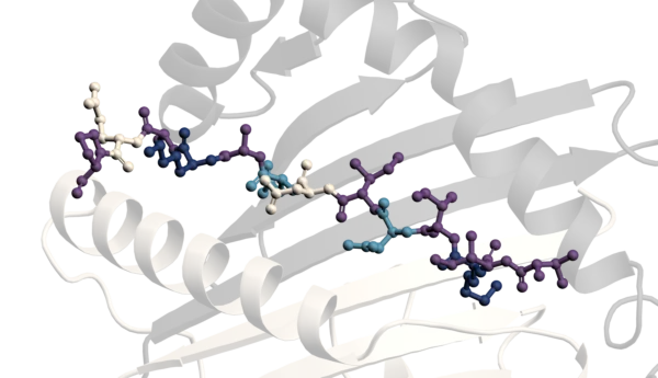

Integrating FIDA™ and Biacore™ SPR for optimal results.


Discover precise insights into brain neurochemistry with Sygnature Discovery's in vivo microdialysis and cOFM services. With over 20 years of expertise, we design bespoke studies that reveal how compounds modulate neurotransmitter systems in health and disease. Using UHPLC/HPLC with electrochemical detection or mass spectrometry, we deliver robust PK/PD data to support confident CNS decision making.


July 30, 2015
Inobrodib, an exciting, first-in-class oral anti-cancer drug in clinical development by CellCentric, was collaboratively designed, synthesized and supported on its pre-clinical journey by an integrated project team at Sygnature Discovery. Inobrodib is now showing promising results in Phase I and II trials for multiple myeloma and other cancer types.

February 3, 2026

Stay up to date with the latest company updates, milestones, and announcements.
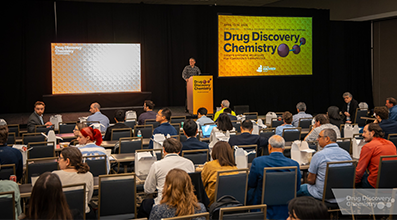
Explore upcoming conferences and scientific meetings where you can connect with our team.

At Sygnature Discovery, we deliver world-leading drug discovery solutions to accelerate your compound from idea to clinic.

Our leadership team brings diverse experience and insight, driving collaboration and innovation across drug discovery.

Explore careers at Sygnature Discovery and join a global team committed to science, collaboration, and integrity.
Inobrodib, an exciting, first-in-class oral anti-cancer drug in clinical development by CellCentric, was collaboratively designed, synthesized and supported on its pre-clinical journey by an integrated project team at Sygnature Discovery. Inobrodib is now showing promising results in Phase I and II trials for multiple myeloma and other cancer types.
At Sygnature Discovery, we deliver world-leading drug discovery solutions to accelerate your compound from idea to clinic.
Our leadership team brings diverse experience and insight, driving collaboration and innovation across drug discovery.
Explore careers at Sygnature Discovery and join a global team committed to science, collaboration, and integrity.
The use of endogenous brain biomarkers to understand how the brain functions in health and disease is key to developing new treatments for neurodevelopmental, neuropsychiatric and neurodegenerative disorders. As translational pharmacodynamic measures, biomarkers enable researchers to monitor disease-relevant biological processes and determine whether experimental treatments are producing the desired functional effects on neural circuits or pathology.
There are many ways in vitro and ex vivo to assess changes in brain biomarkers, but they each have their limitations. For example, in vitro assays often use cells to assess a novel treatment’s effect on a single cell type or receptor. This allows good assessment of target engagement and function at a single cell or receptor level but does not inform on how that interaction affects the broader brain network, which involves diverse cell types operating across a range of interconnected brain regions. Ex vivo tissue analysis can give that broader understanding of how a novel treatment is influencing relevant biomarkers across brain regions. However, its limitation is that it captures only a single snapshot in time when the animal was terminated and tissue collected. This is where in vivo neurochemistry is invaluable as it provides a dynamic temporal measure of biomarker changes.
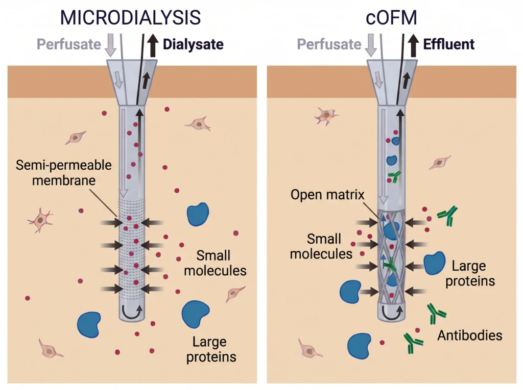
Microdialysis is a long-standing method used to bridge the gap between in vitro and ex vivo methods that allows assessment of the effects of a novel treatment in alive, naturally behaving animals over time. It takes advantage of probes that are most commonly made from a metal shaft with a porous membrane at the tip which contains pores of a specific size that can range from 6kDa to 2MDa. The probes are implanted into the targeted brain region and perfused with artificial cerebrospinal fluid (aCSF), allowing any biomarkers in the interstitial fluid (ISF) around the outside of the membrane and smaller than the membrane pore size to diffuse across and into the aCSF perfusing through the probe. The aCSF containing biomarkers from the brain is collected from the probe’s outlet and can then be used to determine the concentration of the biomarkers of interest in that brain region or regions. This is typically achieved using HPLC-ECD and/or LC-MS/MS. As samples can be continuously collected over time this method gives you a temporal picture of changes to the biomarker of interest that a novel treatment is causing, in a specific brain region from an intact network. It is a powerful disease agnostic method that allows the assessment of functional changes, as a result of the administration of a novel treatment, and assessment of their effectiveness to elicit the changes they are designed to make1. The main group of brain biomarkers that microdialysis is used for are neurotransmitters. Most novel treatments historically developed for CNS disorders have primarily focused on altering neurotransmitter signalling to either enhance where there is a deficit or dampen where there is a surplus. This method has been finely tuned to suit this purpose as it is routinely used in assessing effect on brain function by monitoring changes in relevant neurotransmitter levels; both as a marker that the novel treatment is effectively engaging its target, but also to confirm any potential abuse liability of a new treatment by the assessment of its effect on dopamine release in the nucleus accumbens2,3.
This method, however, still has its limitations. Traditionally pore sizes and the materials the probes and tubing have been made from have precluded its use for measuring large endogenous molecules such as proteins and peptides or highly lipophilic molecules. Over the years this has been, in part, resolved by the development of probes with larger pore size membranes and materials that are less “sticky” for these larger molecules, including by adding small amounts of bovine serum albumin protein (BSA) to the aCSF perfusate to help aid recovery.
The other main disadvantage of this method is of particular importance when assessing either blood brain barrier (BBB) penetrance of a novel treatment or looking at disruption of the BBB itself. The fact that the probes are surgically implanted into the brain means the BBB is inevitably damaged in the process. This disruption means that when you are looking at biomarkers that are present in both periphery and brain you cannot be 100% confident that what you are measuring in those samples is entirely from the brain. The same applies to using microdialysis for pharmacodynamic (PD) studies where you are assessing brain penetrance of a novel treatment that has been administered systemically. You can limit this disadvantage by implanting the probes directly and leaving them for up to 2 weeks to allow the blood brain barrier to heal around the probe, however the way microdialysis probes are designed tends to preclude this option. The membrane material tends to cause glial cells to react to its presence and form a glial scar around the membrane blocking the pores and therefore making the probe unusable within 36 hours. They also need constant perfusion once implanted to maintain the flow path integrity, this would mean having animals hooked up to perfusion pump systems for days or weeks on end. Guide cannula can be used to try and address this issue by implanting them and leaving the animals to recover for up to 2 weeks to allow the BBB to heal around the cannula but there will still be a small area of disturbance around the membrane as it is inserted into and exits from the end of the guide cannula and this disturbance is exactly where samples are collected from.
The biggest leap forward in the area of in vivo neurochemistry has been the development of the cerebral open flow microperfusion (cOFM) method by Joanneum Research GmbH. This development focussed on changes to the design of the traditional microdialysis probe to address the disadvantages outlined above. Specifically, they changed the tip from having a membrane to having an open matrix design with diameter of each hole being 100 µm. They also developed these new probes using materials that were inherently low binding for proteins, peptides and other lipophilic analytes and that are inert in tissue so even after long term implantation there is no activation of glial cells and no glial scar formation4,5.
This newer method not only allows easier collection of larger and more lipophilic biomarkers6, but the open matrix design means that the incoming aCSF mixes directly with the ISF and the sample collected from the outlet is the whole ISF diluted in the aCSF being perfused through the probe. This fact and the inert materials the probes are made from leads to these probes having a better recovery rate of biomarkers than traditional microdialysis probes, giving you an advantage when wanting to measure analytes that have inherently low levels. Having the ability to collect a diluted ISF sample also broadens the range of biomarkers that can be measured from the small molecules, such as neurotransmitters, up to the largest such as proteins, antibodies and nanoparticles. The only limitation on what can be measured in these samples is the assays used and their detection limits. Levels in these samples may be higher than in traditional microdialysis samples but will be lower than found in tissue homogenates as it is only collecting what is in the ISF and the levels are diluted by the aCSF perfusate.
Lastly the way they are designed with the probe itself being the guide cannula with a healing dummy insert to seal the probe when not in use means animals can live in normal home cages while recovering and between any sampling sessions. When it comes to sampling from these probes the healing dummy is replaced with a sampling insert with this change over all being internal to the implanted probe leading to no further disturbance or damage to the brain tissue leaving the healed BBB intact5. This is a huge advantage when looking at brain penetrance of a novel treatment or looking at BBB integrity such as in neuroinflammation models7.
This really depends on the question being asked. For studies that are purely looking at the most common neurotransmitters (such as monoamine, GABA, glutamate, ACh and their metabolites), the long history and development of traditional microdialysis probes and the analysis methods such as HPLC-ECD and LC-MS/MS that go along with it are the better option. The filtered nature of the dialysate sample from a microdialysis probe gives a smoother profile to the changes and less variability at each time point, making it ideal to detect subtle changes in the levels of these neurotransmitters which may be needed in PK/PD study designs. However, if you are looking at larger endogenous biomarkers (such as proteins and peptides) or are wanting to assess brain penetrance of larger novel treatments such as oligonucleotides and antibodies, then cOFM is the method of choice. Its open matrix design and low-bind materials make it ideally suited for assessing these new types of biologic-based treatments.
When you work with us, you leverage the extensive knowledge and experience of our scientists. We guide you to use the most appropriate technique to tackle the particular nuances or your target and aims your project. Beyond that we work with you to interpret the data that is generated to inform the next set of experiments, aiming for the most efficient pathway toward the clinic.



Peak Proteins, NuChem Sciences, and SB Drug Discovery have now fully integrated with Sygnature Discovery.