



Featured Resources
-
STORM Therapeutics: From Lead to Pre-Candidate Nomination in 18 Months

STORM Therapeutics: From Lead to Pre-Candidate Nomination in 18 Months
Case study




Discover precise insights into brain neurochemistry with Sygnature Discovery's in vivo microdialysis and cOFM services. With over 20 years of expertise, we design bespoke studies that reveal how compounds modulate neurotransmitter systems in health and disease. Using UHPLC/HPLC with electrochemical detection or mass spectrometry, we deliver robust PK/PD data to support confident CNS decision making.


July 30, 2015

Inobrodib, an exciting, first-in-class oral anti-cancer drug in clinical development by CellCentric, was collaboratively designed, synthesised and supported on its pre-clinical journey by an integrated project team at Sygnature Discovery. Inobrodib is now showing promising results in Phase I and II trials for multiple myeloma and other cancer types.

In drug discovery, generating targets is no longer the challenge. The real question is how to identify the few worth investing months of research and significant resources to pursue. Hear expert perspectives on how AI, pathway analysis and scientific expertise are shaping modern target identification.
July 7, 2026

Stay up to date with the latest company updates, milestones, and announcements.
Explore upcoming conferences and scientific meetings where you can connect with our team.

At Sygnature Discovery, we deliver world-leading drug discovery solutions to accelerate your compound from idea to clinic.

Our leadership team brings diverse experience and insight, driving collaboration and innovation across drug discovery.

Explore careers at Sygnature Discovery and join a global team committed to science, collaboration, and integrity.
Inobrodib, an exciting, first-in-class oral anti-cancer drug in clinical development by CellCentric, was collaboratively designed, synthesised and supported on its pre-clinical journey by an integrated project team at Sygnature Discovery. Inobrodib is now showing promising results in Phase I and II trials for multiple myeloma and other cancer types.
At Sygnature Discovery, we deliver world-leading drug discovery solutions to accelerate your compound from idea to clinic.
Our leadership team brings diverse experience and insight, driving collaboration and innovation across drug discovery.
Explore careers at Sygnature Discovery and join a global team committed to science, collaboration, and integrity.
“The human brain is the only thing on the planet that is trying to understand itself”
from a talk on the NMDA receptor & memory at Zeneca Pharmaceuticals 1992. R. Mott
“Dear Hilde, if the human brain was simple enough for us to understand, we would still be so stupid that we couldn’t understand it”
from Sophie’s World by Jostein Gaarder
Few proteins have had as profound an impact on modern biology as channelrhodopsin. What began as an investigation into how microscopic green algae sense and respond to light ultimately led to one of the most transformative technologies in neuroscience: optogenetics.
Today, channelrhodopsins allow researchers to control the activity of specific neurons with millisecond precision using light, enabling experiments that were previously impossible. The impact of this technology was recognized with the 2026 Nobel Prize for Medicine.
From a protein biochemistry perspective, channelrhodopsins are fascinating molecules. They combine a light-sensing chromophore with an ion channel in a single protein, directly converting photons into electrical signals without requiring intermediary signaling pathways. This elegant molecular design explains both their biological function in algae and their extraordinary utility as research tools to drive our understanding of how the human brain actually works.
It all began with the unicellular green alga Chlamydomonas reinhardtii, a photosynthetic microorganism that can actively swim toward or away from light sources, a behavior known as phototaxis.
Researchers in the 1970s and 1980s sought to understand the molecular basis of this light-guided movement. Physiological studies demonstrated that illumination of the algal eyespot triggered rapid changes in membrane potential and ion fluxes, suggesting the presence of a specialized photoreceptor linked to ion transport.
A major clue emerged from studies of microbial rhodopsins. Bacteriorhodopsin and halorhodopsin had already been identified in archaea as retinal-containing membrane proteins capable of light-driven ion transport. Scientists suspected that a related class of proteins might exist in algae.
The breakthrough came in the early 2000s when genomic and electrophysiological investigations identified two novel photoreceptor proteins in C. reinhardtii:
In 2002, Nagel and colleagues demonstrated that Channelrhodopsin-1 functions as a light-gated ion channel. Shortly thereafter, Channelrhodopsin-2 was shown to mediate rapid light-induced cation conductance. These findings revealed an entirely new type of photoreceptor: a single-component protein that directly converts light into ion flux.
Unlike vertebrate visual rhodopsins, which activate G-protein signaling cascades, channelrhodopsins operate as much simpler, self-contained molecular machines.
The full story of these initial observations, the subsequent studies into the underlying mechanism via the ChR1 and 2 proteins and how the proteins ultimately ended up as the tools powering the engine of Optogenetics is beautifully described in this Nobel Prize announcement article.
It is a wonderful scientific journey, beginning with Hegemann’s curiosity to understand how algae sense light, followed by Nagel’s work identifying the proteins underlying these responses and uncovering the mechanism. The final “creative” moment, demonstrating the power of cross functional collaboration, came when Desseroth and colleagues recognized that channelrhodopsins could be used to trigger electrical impulses in nerve cells. The subsequent development of methods to make this work in living animals laid the foundations for optogenetics.
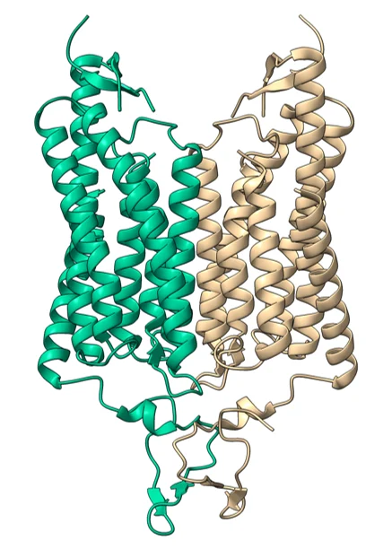
Channelrhodopsins belong to the microbial rhodopsin superfamily. Like other rhodopsins, they contain:
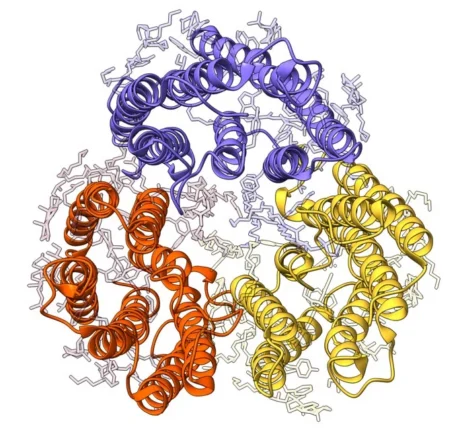
Significant progress in understanding channelrhodopsin function came from structural biology, with the first high-resolution crystal structure of a channelrhodopsin being reported in 2012.
Beyond simply revealing the architecture of the protein, these studies helped explain how absorption of light by retinal is coupled to opening of the ion-conducting pore. As an integral membrane protein, however, channelrhodopsin presented many of the challenges familiar to structural biologists, including recombinant expression, extraction from lipid membranes and maintenance of stability outside its native environment.
To solve this initial structure Kato et al., (2012) screened a number of engineered channelrhodopsin constructs from C. reinhardtii and other organisms. The successful construct was a chimeric version of C. reinhardtii channelrhodopsin that they named C1C2, which consisted of ChR1 without the C terminus, with the last two TM regions exchanged for those from ChR2. At the C-terminus a TEV-eGFP-8His tag was added. The eGFP for use in the initial construct screening in HEK293 cells using fSEC and the His tag to aid purification. To support the crystallization studies this construct was expressed in baculovirus infected Sf+ insect cells and purified following solubilization of the membrane fraction.
This and subsequent structural studies provided direct insights into:
This combination of features makes channelrhodopsin particularly effective at depolarizing cells. In native algae, this depolarization triggers those behavioral responses to light that Hegemann and others had observed.
The central biochemical event underlying channelrhodopsin function is retinal photoisomerization.
In the dark state:
Upon absorption of a photon:
This process occurs on the microsecond timescale, and the resulting open state permits the passage of cations including: H⁺, Na⁺, K⁺ and Ca²⁺. The protein subsequently progresses through a series of photointermediates before returning to its original dark state.
The complete photocycle occurs within milliseconds, allowing repeated stimulation at high frequencies. And it is this rapid response that was one of the key factors that meant that channelrhodopsins were ideal to be developed as tools for use in the new field of optogenetics.
Subsequent cryo-electron microscopy and crystallographic studies on the C. reinhardtii and other related channelrhodopsins have revealed how subtle amino acid substitutions can dramatically alter:
This structural understanding laid the foundation for protein engineering efforts that transformed channelrhodopsins into versatile optogenetic tools. (See later for more detail)
Although the algal proteins themselves were fascinating, their greatest impact came from an unexpected application, described more fully in the Nobel Prize announcement article.
In 2005, Karl Deisseroth, Edward Boyden and colleagues demonstrated that expression of Channelrhodopsin-2 in mammalian neurons allowed neuronal firing to be controlled by pulses of blue light.
This was a revolutionary concept.
Rather than using electrodes to stimulate neurons:
The simplicity of the system was unprecedented because channelrhodopsin required only:
No additional signaling components were necessary.
The technology quickly became known as optogenetics, combining optical control with genetic targeting.
The concept was remarkably simple, but its impact was transformative. For the first time, researchers could selectively activate specific neurons within a living animal and directly observe the effects on behavior. Neuroscience could move beyond observing correlations and begin testing cause and effect.
From a protein science perspective, channelrhodopsin is particularly elegant. Unlike many biological signaling systems that require multiple proteins, channelrhodopsin combines light sensing and ion channel activity within a single seven-transmembrane protein, making it both mechanistically fascinating and highly amenable to experimental study.
Several biochemical properties made channelrhodopsin uniquely suitable.
Single-Component Functionality
Fast Kinetics
Direct Electrical Control
Genetic Targetability
The original Channelrhodopsin-2 molecule was remarkably effective but based on the cryo-EM and X-ray structure data, researchers soon began engineering variants with improved properties, generating an extensive toolbox tailored for specific experimental needs to progress the optogenetics field.
Examples include:
The use of channelrhodopsin tools has branched out to other areas of research. Applications include:
Researchers are also exploring therapeutic applications for:
Channelrhodopsin represents one of the most important discoveries at the intersection of protein biochemistry, structural biology and neuroscience. Originally evolved to help green algae navigate their light environment, this retinal-binding membrane protein revealed a fundamentally new mechanism for converting photons directly into ionic currents.
Its elegant architecture, consisting of a seven-transmembrane rhodopsin scaffold fused to a light-gated ion channel, provided a ready-made molecular switch perfectly suited for biological engineering. The subsequent adoption of Channelrhodopsin-2 as the foundation of optogenetics revolutionized the study of neural circuits and opened entirely new avenues for understanding physiology and disease.
In many ways, channelrhodopsin embodies one of the most powerful ideas in science: that curiosity driven investigations can deliver transformative technologies. A protein that evolved to help a single-celled alga navigate its environment now allows researchers to probe the workings of the most complex biological structure known, the human brain.


Peak Proteins, NuChem Sciences, and SB Drug Discovery have now fully integrated with Sygnature Discovery.