


Featured Resources
-
STORM Therapeutics: From Lead to Pre-Candidate Nomination in 18 Months

STORM Therapeutics: From Lead to Pre-Candidate Nomination in 18 Months
Case study




Discover precise insights into brain neurochemistry with Sygnature Discovery's in vivo microdialysis and cOFM services. With over 20 years of expertise, we design bespoke studies that reveal how compounds modulate neurotransmitter systems in health and disease. Using UHPLC/HPLC with electrochemical detection or mass spectrometry, we deliver robust PK/PD data to support confident CNS decision making.


July 30, 2015

Inobrodib, an exciting, first-in-class oral anti-cancer drug in clinical development by CellCentric, was collaboratively designed, synthesized and supported on its pre-clinical journey by an integrated project team at Sygnature Discovery. Inobrodib is now showing promising results in Phase I and II trials for multiple myeloma and other cancer types.

In drug discovery, generating targets is no longer the challenge. The real question is how to identify the few worth investing months of research and significant resources to pursue. Hear expert perspectives on how AI, pathway analysis and scientific expertise are shaping modern target identification.
July 7, 2026

Stay up to date with the latest company updates, milestones, and announcements.
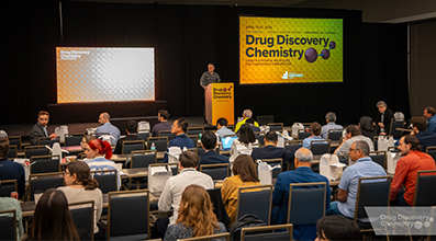
Explore upcoming conferences and scientific meetings where you can connect with our team.

At Sygnature Discovery, we deliver world-leading drug discovery solutions to accelerate your compound from idea to clinic.

Our leadership team brings diverse experience and insight, driving collaboration and innovation across drug discovery.

Explore careers at Sygnature Discovery and join a global team committed to science, collaboration, and integrity.
Inobrodib, an exciting, first-in-class oral anti-cancer drug in clinical development by CellCentric, was collaboratively designed, synthesized and supported on its pre-clinical journey by an integrated project team at Sygnature Discovery. Inobrodib is now showing promising results in Phase I and II trials for multiple myeloma and other cancer types.
At Sygnature Discovery, we deliver world-leading drug discovery solutions to accelerate your compound from idea to clinic.
Our leadership team brings diverse experience and insight, driving collaboration and innovation across drug discovery.
Explore careers at Sygnature Discovery and join a global team committed to science, collaboration, and integrity.
Nuclear Magnetic Resonance (NMR) spectroscopy is one of the most powerful techniques available for studying biomolecules in solution. Unlike many analytical methods, NMR can provide detailed information on protein structure, stability, dynamics, and molecular interactions under near-physiological conditions.
At Sygnature Discovery, we have extensive experience producing isotope-labeled proteins to support NMR studies. Combined with specialist NMR expertise and access to leading NMR facilities, we can support a range of applications from protein quality assessment through to protein-ligand interaction analysis and structural characterization.
NMR exploits the magnetic properties of certain atomic nuclei, including hydrogen (¹H), nitrogen (¹⁵N), and carbon (¹³C). When placed in a strong magnetic field and exposed to radiofrequency pulses, these nuclei generate signals that provide detailed information about their molecular environment.
Since its development in the mid-20th century, advances in superconducting magnet technology and data processing have transformed NMR into a versatile tool for investigating proteins, nucleic acids, carbohydrates, and small molecules.
For many protein-focused applications, isotopically labeled proteins containing ¹⁵N and ¹³C are produced to improve sensitivity and enable residue-specific analysis. These labeled proteins are typically generated through recombinant expression in bacterial systems using isotope-enriched growth media.
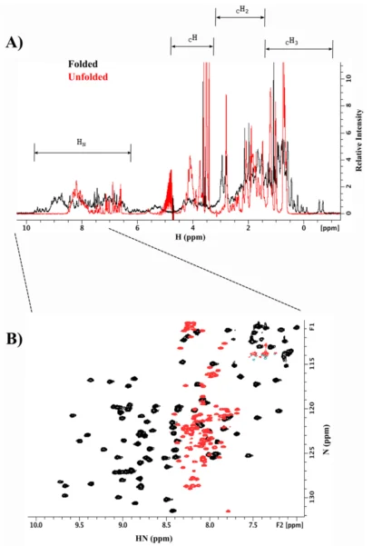
One of the most valuable applications of NMR is the rapid assessment of protein quality.
For unlabeled proteins, one-dimensional (1D) proton NMR spectra can provide information about whether a protein is folded or unfolded. Well-folded proteins produce highly dispersed resonances, whereas unfolded proteins exhibit a much narrower range of signals.
When ¹⁵N-labeled proteins are available, two-dimensional (2D) NMR experiments provide significantly greater detail. These spectra can act as molecular fingerprints, allowing scientists to assess protein integrity, stability, and conformational changes with residue-level resolution.
This capability makes NMR a valuable quality-control tool during protein production and characterization workflows.
NMR is uniquely suited to studying molecular interactions in solution and has become an important technique in drug discovery, particularly for fragment screening and hit validation.
One of its key strengths is the ability to detect interactions across a wide range of affinities while providing atomic-level information about binding events.
Two complementary approaches are commonly used.
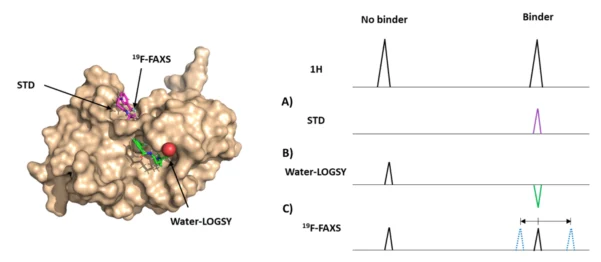
In ligand-observed experiments, the small molecule acts as the reporter molecule.
These methods do not require isotope-labeled protein and can be applied to proteins of virtually any size.
Common techniques include:
These approaches are widely used to identify and confirm binding interactions during early-stage drug discovery and fragment screening campaigns (Figure 2).
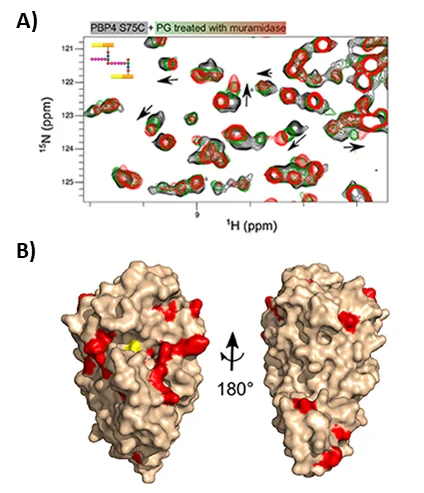
Protein-observed experiments use ¹⁵N- or ¹³C-labeled proteins and provide more detailed information about the interaction (Figure 3).
By monitoring changes in individual resonances during ligand titration experiments, it is possible to:
Importantly, NMR can evaluate interactions with a wide range of binding partners, including small molecules, peptides, proteins, nucleic acids, carbohydrates, and lipids.
NMR provides a unique combination of structural, biophysical, and interaction data that can support multiple stages of a discovery program.
Applications include:
By revealing how proteins behave and interact in solution, NMR can help researchers gain confidence in candidate molecules and make better-informed decisions during drug discovery.
NMR remains one of the most versatile techniques available for characterizing proteins and molecular interactions. Whether assessing protein quality, validating ligand binding, or investigating molecular mechanisms, NMR provides insights that are often difficult to obtain through other analytical approaches.
Combined with expertise in recombinant protein production and isotope labeling, NMR can play a valuable role in addressing challenging protein science and drug discovery questions.

Peak Proteins, NuChem Sciences, and SB Drug Discovery have now fully integrated with Sygnature Discovery.