



Featured Resources
-
STORM Therapeutics: From Lead to Pre-Candidate Nomination in 18 Months

STORM Therapeutics: From Lead to Pre-Candidate Nomination in 18 Months
Case study




Discover precise insights into brain neurochemistry with Sygnature Discovery's in vivo microdialysis and cOFM services. With over 20 years of expertise, we design bespoke studies that reveal how compounds modulate neurotransmitter systems in health and disease. Using UHPLC/HPLC with electrochemical detection or mass spectrometry, we deliver robust PK/PD data to support confident CNS decision making.


July 30, 2015

Inobrodib, an exciting, first-in-class oral anti-cancer drug in clinical development by CellCentric, was collaboratively designed, synthesized and supported on its pre-clinical journey by an integrated project team at Sygnature Discovery. Inobrodib is now showing promising results in Phase I and II trials for multiple myeloma and other cancer types.

In drug discovery, generating targets is no longer the challenge. The real question is how to identify the few worth investing months of research and significant resources to pursue. Hear expert perspectives on how AI, pathway analysis and scientific expertise are shaping modern target identification.
July 7, 2026

Stay up to date with the latest company updates, milestones, and announcements.
Explore upcoming conferences and scientific meetings where you can connect with our team.

At Sygnature Discovery, we deliver world-leading drug discovery solutions to accelerate your compound from idea to clinic.

Our leadership team brings diverse experience and insight, driving collaboration and innovation across drug discovery.

Explore careers at Sygnature Discovery and join a global team committed to science, collaboration, and integrity.
Inobrodib, an exciting, first-in-class oral anti-cancer drug in clinical development by CellCentric, was collaboratively designed, synthesized and supported on its pre-clinical journey by an integrated project team at Sygnature Discovery. Inobrodib is now showing promising results in Phase I and II trials for multiple myeloma and other cancer types.
At Sygnature Discovery, we deliver world-leading drug discovery solutions to accelerate your compound from idea to clinic.
Our leadership team brings diverse experience and insight, driving collaboration and innovation across drug discovery.
Explore careers at Sygnature Discovery and join a global team committed to science, collaboration, and integrity.
The ability to determine and visualize the atomic structure of proteins is invaluable in modern drug discovery. Understanding how proteins are arranged in three dimensions not only helps researchers investigate biological function but also supports the rational design of new therapeutic molecules that can interact with specific targets.
One of the most widely used techniques for determining protein structures is X-ray crystallography. However, obtaining a protein structure is far from a single-step process. Success depends on two critical stages: generating high-quality protein crystals and extracting meaningful information from the resulting X-ray diffraction data. Both stages require specialist expertise and, often, a great deal of patience.
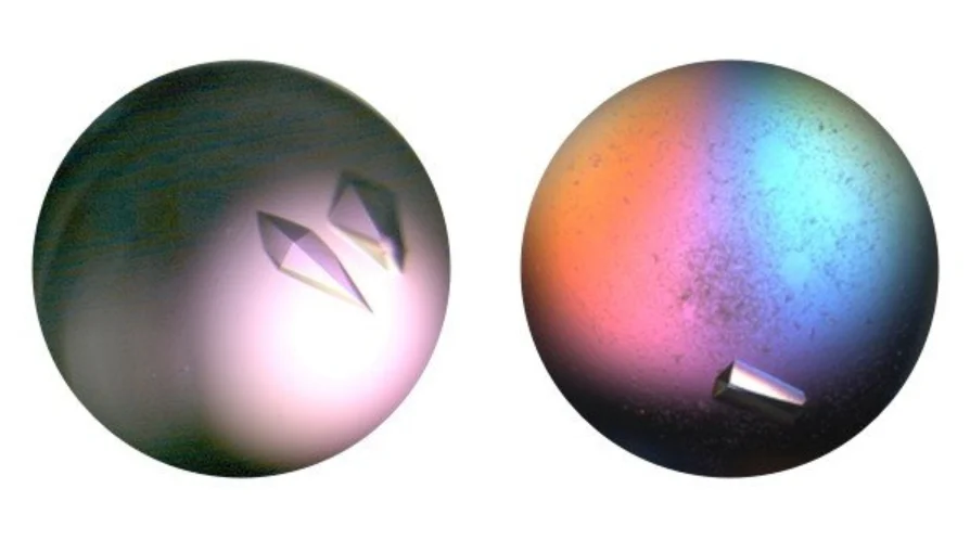
As its name suggests, X-ray crystallography relies on the availability of well-ordered three-dimensional crystals of the target protein. For many projects, producing these crystals is the rate-limiting step in the entire structure determination process.
Unlike other experimental techniques, there is currently no reliable way to predict the optimal crystallization conditions for a protein based solely on its amino acid sequence. Instead, crystallization remains an empirical process that relies on screening large numbers of conditions and systematically optimizing promising hits. This trial-and-error approach has historically given protein crystallization a reputation as something of a “black art” among non-specialists.
Researchers may screen thousands of different crystallization conditions while varying factors such as pH, temperature, salts, precipitants, and protein concentration in an effort to identify conditions that produce suitable crystals. When successful, these crystals provide the highly ordered molecular arrangement required for X-ray diffraction experiments.
Although extensive literature exists on protein crystallization methodologies there remains a need for accessible introductions aimed at scientists who may be considering structural studies for the first time.
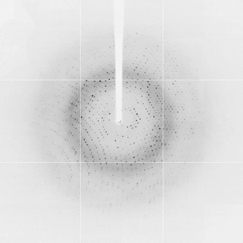
Obtaining protein crystals is only the beginning of the structure determination process. Once suitable crystals have been generated, they are exposed to an X-ray beam to produce diffraction images.
When X-rays interact with the highly ordered lattice of protein molecules within the crystal, they generate a distinctive diffraction pattern that contains information about the arrangement of atoms within the protein.
The quality of these diffraction images is heavily dependent on crystal quality. Consequently, the significant time and resources invested in identifying optimal crystallization conditions are justified by the potential to generate higher-resolution structural information.
However, diffraction images alone do not provide an immediately interpretable protein structure. The raw experimental data must first undergo a series of computational processing steps before it can be used for structure determination and model building.
Given the effort required to obtain suitable crystals, it is important to extract as much information as possible from every diffraction experiment.
Although most synchrotron facilities now perform initial data processing automatically, manual intervention is often still required. This is particularly true for newly obtained crystal forms where there may be uncertainty regarding the correct space group or when more complex crystallographic phenomena, such as crystal twinning, are present.
For this reason, even non-experts can benefit from developing a basic understanding of how X-ray diffraction data are processed.
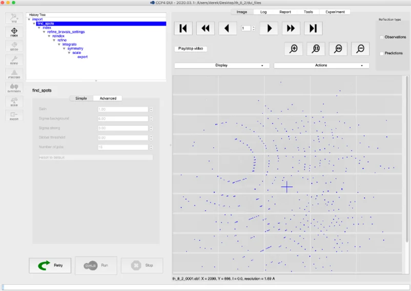
Today’s crystallographers have access to several powerful software packages capable of processing diffraction data efficiently and accurately.
Among the most widely used are:
These programs perform a range of computational tasks, including:
Each stage contributes to the creation of a refined dataset that accurately represents the experimental observations and can be used for subsequent structure solution and model refinement.
Protein crystallization and X-ray data processing are often viewed as separate disciplines, but they are fundamentally linked.
High-quality crystals are essential for producing high-quality diffraction data, while sophisticated data processing is required to maximize the value of that experimental data. Weaknesses at either stage can limit the quality of the final structural model.
Together, these techniques form the foundation of X-ray crystallography and enable researchers to answer key questions about protein function, ligand binding, and molecular mechanisms of action.
Protein crystallization and X-ray data processing are often viewed as separate technical disciplines, but in practice they are interconnected steps within a broader structural biology workflow. Success depends on expertise at every stage, from designing the right protein construct and identifying crystallization conditions to collecting high-quality diffraction data and generating robust structural models.
Sygnature Discovery’s Protein Sciences and Structural Biology teams provide integrated support across this entire process. Our scientists have extensive experience tackling challenging targets, developing crystallization strategies, processing complex datasets, and delivering high-resolution structures that support decision-making across drug discovery programs.
By combining specialist structural biology expertise with Sygnature Discovery’s wider drug discovery capabilities, we help clients move beyond simply generating structures to understanding how those structures can be used to accelerate medicinal chemistry, reduce project risk, and advance promising programs towards the clinic.

Peak Proteins, NuChem Sciences, and SB Drug Discovery have now fully integrated with Sygnature Discovery.