


Featured Resources
-
STORM Therapeutics: From Lead to Pre-Candidate Nomination in 18 Months

STORM Therapeutics: From Lead to Pre-Candidate Nomination in 18 Months
Case study




Discover precise insights into brain neurochemistry with Sygnature Discovery's in vivo microdialysis and cOFM services. With over 20 years of expertise, we design bespoke studies that reveal how compounds modulate neurotransmitter systems in health and disease. Using UHPLC/HPLC with electrochemical detection or mass spectrometry, we deliver robust PK/PD data to support confident CNS decision making.


July 30, 2015

Inobrodib, an exciting, first-in-class oral anti-cancer drug in clinical development by CellCentric, was collaboratively designed, synthesised and supported on its pre-clinical journey by an integrated project team at Sygnature Discovery. Inobrodib is now showing promising results in Phase I and II trials for multiple myeloma and other cancer types.

In drug discovery, generating targets is no longer the challenge. The real question is how to identify the few worth investing months of research and significant resources to pursue. Hear expert perspectives on how AI, pathway analysis and scientific expertise are shaping modern target identification.
July 7, 2026

Stay up to date with the latest company updates, milestones, and announcements.
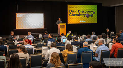
Explore upcoming conferences and scientific meetings where you can connect with our team.

At Sygnature Discovery, we deliver world-leading drug discovery solutions to accelerate your compound from idea to clinic.

Our leadership team brings diverse experience and insight, driving collaboration and innovation across drug discovery.

Explore careers at Sygnature Discovery and join a global team committed to science, collaboration, and integrity.
Inobrodib, an exciting, first-in-class oral anti-cancer drug in clinical development by CellCentric, was collaboratively designed, synthesised and supported on its pre-clinical journey by an integrated project team at Sygnature Discovery. Inobrodib is now showing promising results in Phase I and II trials for multiple myeloma and other cancer types.
At Sygnature Discovery, we deliver world-leading drug discovery solutions to accelerate your compound from idea to clinic.
Our leadership team brings diverse experience and insight, driving collaboration and innovation across drug discovery.
Explore careers at Sygnature Discovery and join a global team committed to science, collaboration, and integrity.
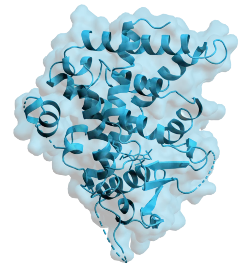
The human Pregnane X Receptor (PXR) is a ligand activated nuclear receptor that plays a central role in regulating xenobiotic metabolism. It functions as a master transcriptional regulator of genes involved in drug metabolism and clearance, including key cytochrome P450 enzymes such as CYP3A4 (1-3).
PXR is characterised by a large and highly flexible ligand binding domain (LBD) enabling the recognition of a wide range of structurally and chemically diverse compounds (4-5). Consequently, PXR is involved in the initial phases of drug metabolism but can also contribute to undesirable effects such as drug–drug interactions, reduced efficacy of anticancer treatments, and the accumulation of toxic metabolites (6). As a result, PXR has emerged as an important target in pharmaceutical research, with ongoing efforts aimed at improving drug efficacy and guiding the development of new therapeutic strategies.
Whilst the intrinsic flexibility of PXR underpins its biological function, it also presents challenges for structural biology. In particular, the conformational heterogeneity of the LBD can hinder crystallisation. To overcome this, we expressed the human PXR ligand binding domain (residues 130 – 434) fused to a stabilising coactivator peptide derived from SRC-1. This strategy, widely reported in the literature, promotes conformational stabilisation of the receptor by mimicking coactivator binding, thereby facilitating crystallisation and structural characterisation (1).
The PXR construct was successfully expressed in E. coli using TB autoinduction media, yielding soluble protein suitable for downstream purification.
Purification was then performed using a twostep workflow:
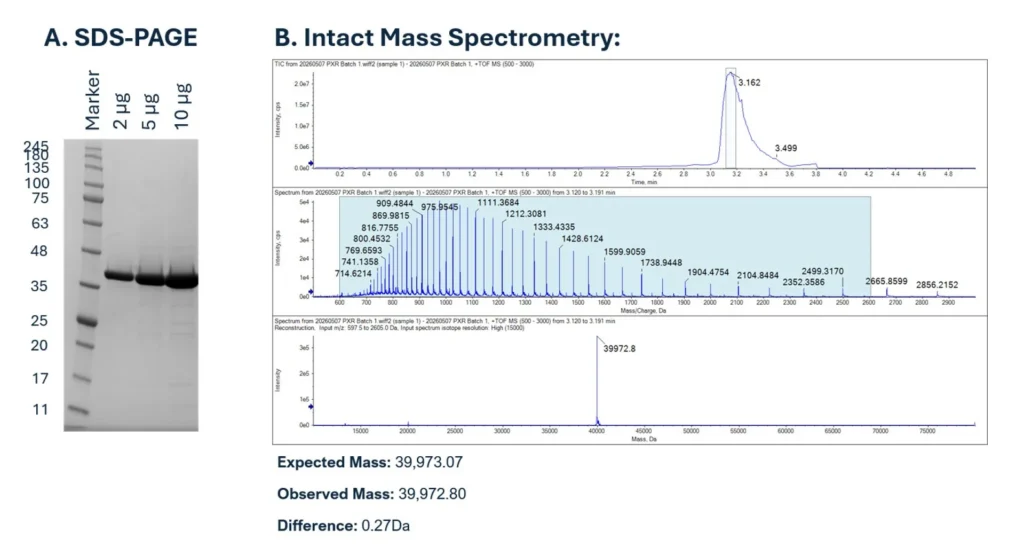
Following IMAC, SDS-PAGE analysis confirmed the presence of the target protein at ~40 kDa, consistent with the expected molecular weight. To achieve the high level of purity and homogeneity required for crystallography, the IMAC eluate was further purified by size exclusion chromatography (SEC). SEC successfully separated PXR from the higher molecular weight contaminants, producing a well defined main peak corresponding to monomeric PXR. Stringent pooling of fractions was performed to maximise homogeneity, which is essential for crystallography. The final protein batch was highly pure (Figure 1A) and showed excellent agreement between expected and observed molecular mass by mass spectrometry (Δ0.27 Da) confirming sample identity (Figure 1B).
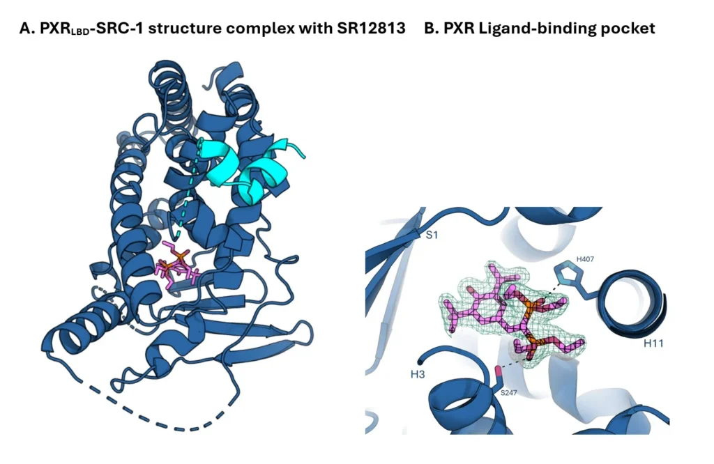
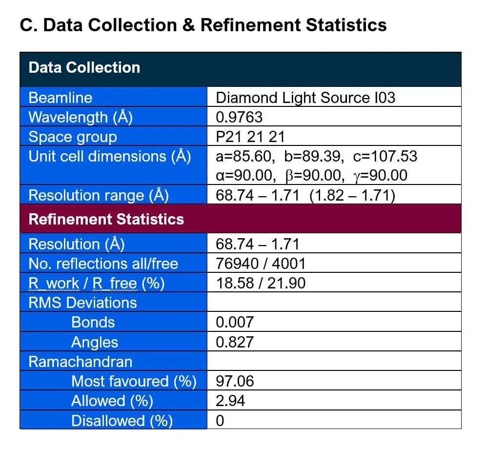
With the purified PXR fused to the SRC-1 peptide, broad and literature informed crystallisation trials were initiated employing a combination of sitting and hanging drop vapour diffusion methods. The presence of the SRC-1 co-activator peptide was key to stabilising the receptor in an active conformation and reduced the conformational heterogeneity which in turn increased the likelihood of crystal formation, an approach consistent with previously reported PXR structures. The final co-crystal structure, in complex with the cholesterol-lowering agent SR12813, belonged to the space group P212121 and was solved at 1.71 Å resolution using data collected at Diamond Light Source and processed with STARANISO (Figure 2).
The resulting crystal structure provides valuable insights into the architecture of the PXR ligand binding domain and establishes a robust platform for future ligand bound co-crystallisation studies. Importantly, this system enables the screening of client supplied compounds directly against PXR, allowing rapid assessment of binding and structural information to be determined on ligand receptor interactions, binding modes, and key contact residues.
Crucially, our integrated teams at Sygnature Discovery can provide an iterative, structure guided design process for client’s compounds. Following initial screening and co-crystallisation, with PXR or other protein targets of interest, ligand bound structures can guide rational optimisation of compounds to reduce or prevent interactions with PXR and correspondingly guide modifications to improve target protein interactions and hence potency. By combining structural biology with medicinal chemistry expertise in-house, this approach allows rapid, coordinated refinement of compounds based on direct experimental insights.
Together, this platform provides a powerful structure guided approach for compound optimisation, enabling clients to refine chemical scaffolds based on direct experimental evidence and ultimately accelerate the development of safer and more effective therapeutics.
This work demonstrates our ability to deliver high quality structural biology solutions for challenging targets such as nuclear receptors. Key highlights include:
With its central role in drug metabolism, PXR remains a critical target in drug discovery. Our expertise enables rapid progression from construct design to structural insight, helping clients better understand ligand binding and guide medicinal chemistry efforts.
Whether you’re targeting PXR or another challenging protein, our gene-to-structure workflow and expert team are ready to support your discovery journey.
Get in touch to explore how Sygnature Discovery can accelerate your structural biology goals.
References
Peak Proteins, NuChem Sciences, and SB Drug Discovery have now fully integrated with Sygnature Discovery.