



Featured Resources
-
STORM Therapeutics: From Lead to Pre-Candidate Nomination in 18 Months

STORM Therapeutics: From Lead to Pre-Candidate Nomination in 18 Months
Case study
📣From lead to pre-candidate nomination in 18 months. Explore the STORM Therapeutics case study




Discover precise insights into brain neurochemistry with Sygnature Discovery's in vivo microdialysis and cOFM services. With over 20 years of expertise, we design bespoke studies that reveal how compounds modulate neurotransmitter systems in health and disease. Using UHPLC/HPLC with electrochemical detection or mass spectrometry, we deliver robust PK/PD data to support confident CNS decision making.


July 30, 2015

Inobrodib, an exciting, first-in-class oral anti-cancer drug in clinical development by CellCentric, was collaboratively designed, synthesised and supported on its pre-clinical journey by an integrated project team at Sygnature Discovery. Inobrodib is now showing promising results in Phase I and II trials for multiple myeloma and other cancer types.

In drug discovery, generating targets is no longer the challenge. The real question is how to identify the few worth investing months of research and significant resources to pursue. Hear expert perspectives on how AI, pathway analysis and scientific expertise are shaping modern target identification.
July 7, 2026

Stay up to date with the latest company updates, milestones, and announcements.
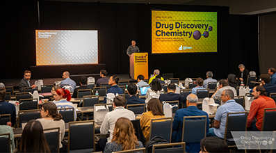
Explore upcoming conferences and scientific meetings where you can connect with our team.

At Sygnature Discovery, we deliver world-leading drug discovery solutions to accelerate your compound from idea to clinic.

Our leadership team brings diverse experience and insight, driving collaboration and innovation across drug discovery.

Explore careers at Sygnature Discovery and join a global team committed to science, collaboration, and integrity.
Inobrodib, an exciting, first-in-class oral anti-cancer drug in clinical development by CellCentric, was collaboratively designed, synthesised and supported on its pre-clinical journey by an integrated project team at Sygnature Discovery. Inobrodib is now showing promising results in Phase I and II trials for multiple myeloma and other cancer types.
At Sygnature Discovery, we deliver world-leading drug discovery solutions to accelerate your compound from idea to clinic.
Our leadership team brings diverse experience and insight, driving collaboration and innovation across drug discovery.
Explore careers at Sygnature Discovery and join a global team committed to science, collaboration, and integrity.
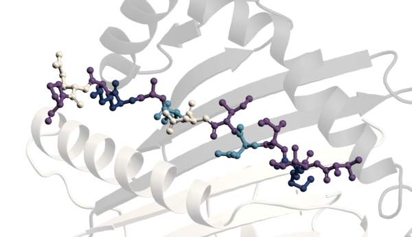
X-ray crystallography is frequently used during drug discovery to determine the molecular details of binding between a target protein and a ligand. Understanding how a ligand interacts with its target can help guide the modification of atoms and functional groups to improve potency and other chemical or biological properties.
There are two common ways to introduce a ligand into a crystal structure: by crystallizing a pre-formed protein-ligand complex or by soaking an apo crystal in a ligand solution after crystallization has taken place. There is no universal answer as to which method should be used, as the choice is highly dependent on both the protein and the ligand. Ligand solubility and potency can have a significant influence on experimental design.
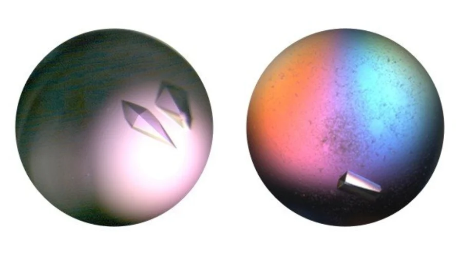
A soakable crystal system can provide a higher-throughput approach when multiple ligands need to be evaluated and requires less protein than co-crystallization approaches. However, it depends on the ability to reproducibly grow apo crystals that can be transferred into soaking solutions without damage.
In practical terms, this means crystallization conditions should not rely on excessive volatile components or solutions that are close to saturation. Crystals also need to be sufficiently robust to allow handling and transfer. Larger three-dimensional crystals are generally easier to work with than very thin plates or needles.
The crystals must usually be able to tolerate at least a few percent Dimethyl Sulfoxide (DMSO), allowing ligands to be added at a suitable excess over the protein concentration. The potency of the ligand also affects the amount required. For potent compounds, ligand occupancy can be achieved at concentrations similar to that of the protein. Compounds with higher dissociation constants generally require a greater excess of ligand. In some cases, ligand solubility limits what can be achieved.
Soaking times can range from minutes to several days. Following transfer into the soaking solution, crystal behaviour can vary considerably. Some crystals remain stable, while others gradually deteriorate or break apart.
When a robust soaking system is available, it is possible to obtain large numbers of ligand-bound structures from relatively small quantities of protein.
In co-crystallization experiments, the protein-ligand complex is formed before crystallization trials are set up. Incubation times can range from approximately 30 minutes to several days.
This approach can be particularly useful when ligands have limited solubility. Complex formation can take place at lower protein concentrations before the sample is concentrated for crystallization, allowing ligands to be present in suitable excess over the protein.
In some situations, additional solvents such as PEG400 or alcohols, particularly diols, can help increase ligand solubility and may be used alone or in combination with DMSO. Similar approaches can also be applied during soaking experiments.
Co-crystallization may also be preferred when proteins do not remain stable in the absence of a ligand.
Many proteins are only stable in solution when complexed with cofactors or nucleotides. Without a stabilizing ligand, proteins may fail to fold correctly, aggregate, or prove difficult to purify to homogeneity. This can limit the number of proteins that can be crystallized as an apo form suitable for soaking experiments.
In other cases, an apo protein may be stable and crystallize readily, but ligand binding causes a conformational change. Under these circumstances, soaking may be unsuccessful and crystal damage can occur.
An example described for the kinase JAK2 demonstrates the role that ligands can play in protein stabilization. Purification of apo JAK2 produced protein that was highly prone to aggregation. However, purification in the presence of staurosporine, a potent kinase inhibitor, generated a stable protein-ligand complex that could be purified and used for crystallization experiments.
These crystals could subsequently be soaked with ligands of interest, although only compounds with a higher affinity for the protein than staurosporine could successfully replace the bound inhibitor.
Proteins do not always behave as expected during purification and crystallization. Some proteins retain their natural ligands throughout purification, while others may acquire ligands from buffers, cryoprotectants, or other reagents used during sample preparation.
It is also important to maintain an appropriate concentration of ligand during cryoprotection. If ligand concentrations are reduced at this stage, weakly bound ligands may dissociate and be lost from the protein before data collection.
Both crystal soaking and co-crystallization are established approaches for generating protein-ligand complex structures. The most suitable method depends on factors including protein stability, ligand potency, ligand solubility, and the ability of crystals to tolerate ligand introduction.
In some cases, ligands serve an additional role by stabilizing proteins during purification and crystallization, permitting structures to be obtained that may otherwise be difficult to generate. Selecting the appropriate strategy and understanding the behaviour of both the protein and ligand are important considerations when pursuing protein-ligand complex structures for drug discovery.

Peak Proteins, NuChem Sciences, and SB Drug Discovery have now fully integrated with Sygnature Discovery.