



Featured Resources
-
STORM Therapeutics: From Lead to Pre-Candidate Nomination in 18 Months

STORM Therapeutics: From Lead to Pre-Candidate Nomination in 18 Months
Case study




Discover precise insights into brain neurochemistry with Sygnature Discovery's in vivo microdialysis and cOFM services. With over 20 years of expertise, we design bespoke studies that reveal how compounds modulate neurotransmitter systems in health and disease. Using UHPLC/HPLC with electrochemical detection or mass spectrometry, we deliver robust PK/PD data to support confident CNS decision making.


July 30, 2015

Inobrodib, an exciting, first-in-class oral anti-cancer drug in clinical development by CellCentric, was collaboratively designed, synthesized and supported on its pre-clinical journey by an integrated project team at Sygnature Discovery. Inobrodib is now showing promising results in Phase I and II trials for multiple myeloma and other cancer types.

In drug discovery, generating targets is no longer the challenge. The real question is how to identify the few worth investing months of research and significant resources to pursue. Hear expert perspectives on how AI, pathway analysis and scientific expertise are shaping modern target identification.
July 7, 2026

Stay up to date with the latest company updates, milestones, and announcements.
Explore upcoming conferences and scientific meetings where you can connect with our team.

At Sygnature Discovery, we deliver world-leading drug discovery solutions to accelerate your compound from idea to clinic.

Our leadership team brings diverse experience and insight, driving collaboration and innovation across drug discovery.

Explore careers at Sygnature Discovery and join a global team committed to science, collaboration, and integrity.
Inobrodib, an exciting, first-in-class oral anti-cancer drug in clinical development by CellCentric, was collaboratively designed, synthesized and supported on its pre-clinical journey by an integrated project team at Sygnature Discovery. Inobrodib is now showing promising results in Phase I and II trials for multiple myeloma and other cancer types.
At Sygnature Discovery, we deliver world-leading drug discovery solutions to accelerate your compound from idea to clinic.
Our leadership team brings diverse experience and insight, driving collaboration and innovation across drug discovery.
Explore careers at Sygnature Discovery and join a global team committed to science, collaboration, and integrity.
Protein stability is a critical factor in many areas of drug discovery, influencing protein production, characterization, structural biology, and ligand screening. While thermal melting experiments are often used as a routine quality control tool, they can also provide valuable insights into protein behavior, buffer optimization, ligand binding, and protein function.
At Sygnature Discovery, thermal stability measurements are routinely used to assess protein quality and support downstream applications such as protein characterization and crystallization studies. Beyond simply determining melting temperature, these assays can provide a deeper understanding of how proteins respond to changes in their environment and interact with potential binding partners.
Several approaches exist for measuring protein thermal stability, but one of the most widely used is the thermofluor stability assay.
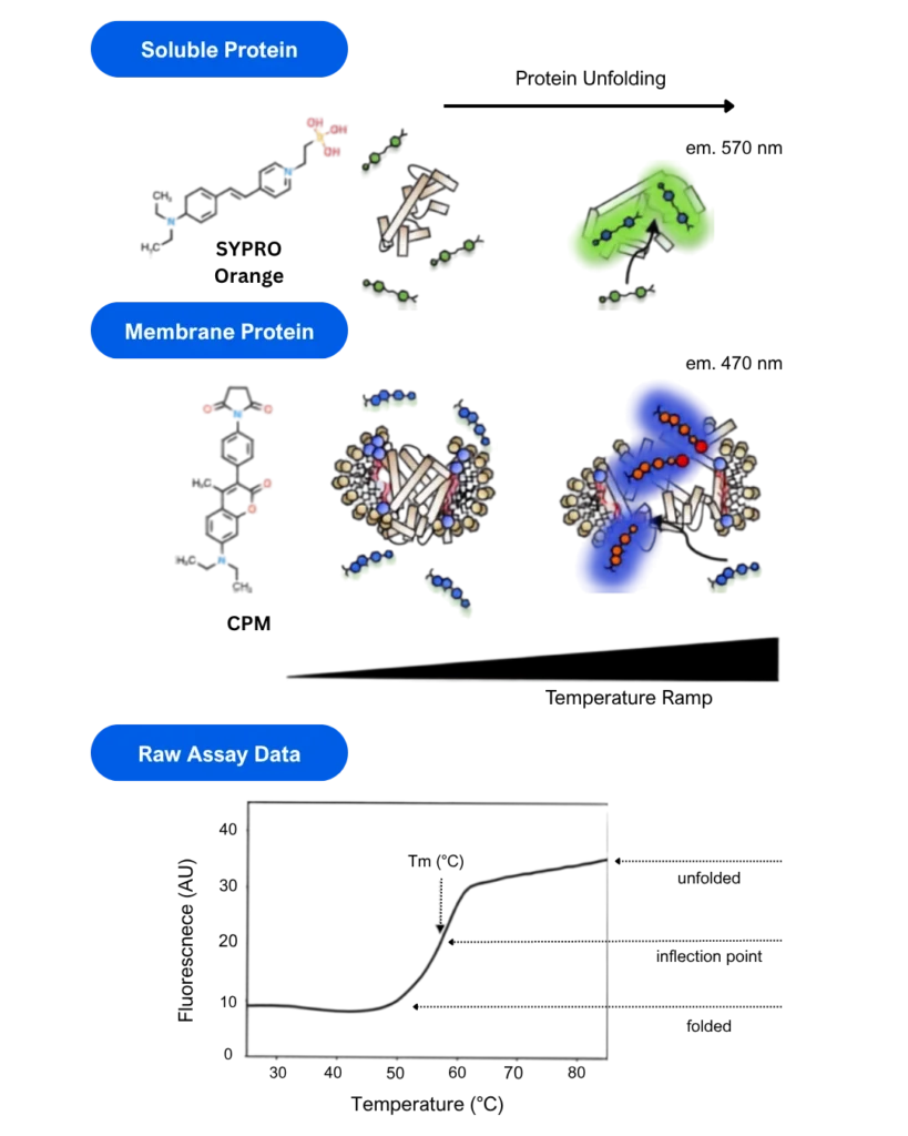
The assay uses environmentally sensitive fluorescent dyes that interact with proteins during thermal unfolding. Two commonly used dyes are:
When these dyes interact with proteins, their fluorescence increases significantly. By monitoring fluorescence while gradually increasing the temperature, it is possible to generate a protein unfolding curve and determine the apparent melting temperature (Tm).
The Tm is commonly used as a relative measure of protein stability under a given set of conditions.
SYPRO Orange is typically used for soluble proteins, while CPM is often preferred for membrane proteins due to the incompatibility of SYPRO Orange with detergent micelles.
The true power of thermofluor stability assays lies not only in measuring Tm values, but also in comparing protein stability under different conditions.
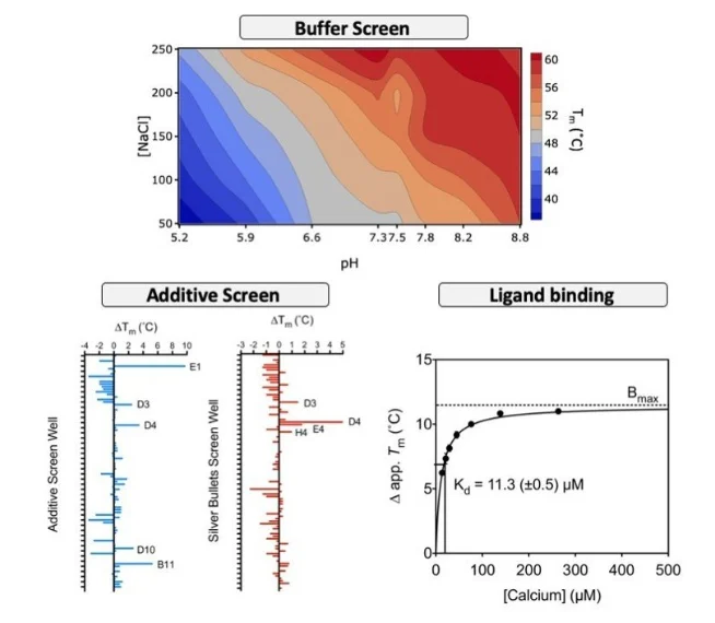
Changes in melting temperature (ΔTm) can be used to assess how factors such as buffer composition, salt concentration, or ligand binding affect protein stability. This approach is commonly referred to as Differential Scanning Fluorimetry (DSF).
By evaluating thermostability shifts between conditions, researchers can obtain information that extends far beyond a simple measure of thermal denaturation.
One common application of DSF is the optimization of protein storage conditions.
Buffer pH and ionic strength can have significant effects on protein stability, and their influence is often interdependent. By systematically varying parameters such as pH and salt concentration, it is possible to identify conditions that maximize protein stability.
This approach can help support protein production workflows by identifying buffer conditions suitable for storage, characterization, and structural studies.
Thermostability shift assays can also be used to assess the effect of ligands on protein stability.
In many cases, binding of a specific ligand stabilizes the protein and results in an increase in melting temperature. This approach can be applied to a wide range of molecule types, including fragments and lead compounds.
Thermofluor assays are frequently used during crystallization optimization to identify additives that improve protein stability. When implemented in high-throughput formats, DSF can also support drug discovery programs by rapidly assessing ligand-induced stabilization.
In some cases, ligand titration experiments can provide a proxy measure of binding affinity.
An example of ligand-induced stabilization following calcium titration is shown in Figure 2.
Thermostability assays can also provide insights into protein function.
Comparing the thermal stability of wild-type and mutant proteins may reveal the importance of specific residues involved in protein activity or mechanism. Changes in stability can help identify regions that contribute to structural integrity, ligand binding, or catalytic function.
As a result, thermofluor assays can support both protein characterization and mechanistic studies.
Thermofluor stability assays are widely used to determine protein melting temperatures and assess protein quality. However, their value extends well beyond routine quality control.
By measuring changes in protein stability under different conditions, differential scanning fluorimetry can support buffer optimization, ligand screening, crystallization studies, and the investigation of protein function. These capabilities make thermofluor assays a versatile tool within modern protein science and drug discovery workflows.




Peak Proteins, NuChem Sciences, and SB Drug Discovery have now fully integrated with Sygnature Discovery.