

Featured Resources
-
STORM Therapeutics: From Lead to Pre-Candidate Nomination in 18 Months

STORM Therapeutics: From Lead to Pre-Candidate Nomination in 18 Months
Case study
📣From lead to pre-candidate nomination in 18 months. Explore the STORM Therapeutics case study


Discover precise insights into brain neurochemistry with Sygnature Discovery's in vivo microdialysis and cOFM services. With over 20 years of expertise, we design bespoke studies that reveal how compounds modulate neurotransmitter systems in health and disease. Using UHPLC/HPLC with electrochemical detection or mass spectrometry, we deliver robust PK/PD data to support confident CNS decision making.


July 30, 2015

Inobrodib, an exciting, first-in-class oral anti-cancer drug in clinical development by CellCentric, was collaboratively designed, synthesised and supported on its pre-clinical journey by an integrated project team at Sygnature Discovery. Inobrodib is now showing promising results in Phase I and II trials for multiple myeloma and other cancer types.

At Sygnature Discovery, we deliver world-leading drug discovery solutions to accelerate your compound from idea to clinic.

Our leadership team brings diverse experience and insight, driving collaboration and innovation across drug discovery.

Explore careers at Sygnature Discovery and join a global team committed to science, collaboration, and integrity.
Inobrodib, an exciting, first-in-class oral anti-cancer drug in clinical development by CellCentric, was collaboratively designed, synthesised and supported on its pre-clinical journey by an integrated project team at Sygnature Discovery. Inobrodib is now showing promising results in Phase I and II trials for multiple myeloma and other cancer types.
At Sygnature Discovery, we deliver world-leading drug discovery solutions to accelerate your compound from idea to clinic.
Our leadership team brings diverse experience and insight, driving collaboration and innovation across drug discovery.
Explore careers at Sygnature Discovery and join a global team committed to science, collaboration, and integrity.
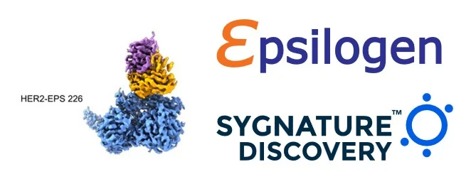
Therapeutic antibodies of the IgE class are attracting increasing interest as a new modality in oncology. In a recent Nature Scientific Reports publication, clinical stage oncology company Epsilogen explores whether increasing monovalent affinity translates into improved anti‑tumour potency for a biotherapeutic IgE.
To address this, Epsilogen engineered a panel of affinity‑matured HER2‑targeting IgE variants and evaluated their biological activity across multiple in vitro and in vivo models. A key part of this study required the generation of high-resolution structural evidence to determine whether increased potency arose from affinity enhancement alone, rather than from changes in epitope specificity.
Using cryo‑EM datasets generated by Epsilogen, in collaboration with the Cambridge cryo‑EM facility, Sygnature Discovery supported the project at the structure elucidation and analysis stage. Our cryo-EM team processed the data, reconstructed 3D maps for the Fab complexes from Epsilogen’s clinical candidates HER2–EPS 226 and HER2–EPS 232, and built and refined atomic models to publication quality.
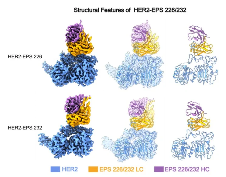
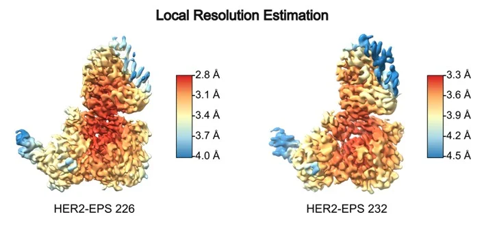
The resulting structures, resolved to 2.98 Å (HER2-EPS 226) and 3.5 Å (HER2-EPS 232) global resolution, enabled detailed definition of the Fab–HER2 interface.
Despite pronounced preferred orientation, a commonly encountered issue in cryo-EM samples, sufficient density was obtained to build accurate models. Local resolution analysis showed that the central binding region was particularly well resolved. This level of detail allowed confident placement of side chains at the epitope–paratope interface and meaningful comparison between variants.
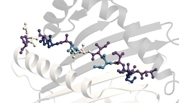
Structural overlay of the two complexes revealed that EPS 226 and the affinity matured variant EPS 232 engage HER2 through conserved CDR mediated interactions at the same epitope.
Figure 3. Structural overlay showing conserved CDR‑mediated recognition of the same HER2 epitope by EPS226 and EPS232 through closely related CDR interactions.
Crucially, this demonstrates that the enhanced biological potency of EPS 232 is driven by increased affinity rather than epitope drift. Together with the biological data, these structures provide definitive evidence that affinity maturation alone can deliver functional improvement in HER2 targeting IgE antibodies.
This project highlights Sygnature’s ability to integrate seamlessly into client workflows. While we offer a full gene to structure cryo-EM pipeline, here we worked from client generated data to deliver high impact structural insight. Our cloud-enabled infrastructure, including scalable cryo-EM data processing on AWS, supports efficient and reproducible handling of large datasets.
For emerging modalities such as therapeutic IgE, structural biology is a powerful tool for derisking candidates, validating mechanisms of action, and informing discovery decisions. We were delighted to support Epsilogen in advancing this important work and contributing to the growing understanding of IgE as a therapeutic platform.
Coordinate models and cryo-EM maps have been deposited in the PDB/EMDB under accession codes 9T3S and EMD55518 (HER2–EPS 226 Fab) and 9T3R and EMD55517 (HER2–EPS 232 Fab).

Peak Proteins, NuChem Sciences, and SB Drug Discovery have now fully integrated with Sygnature Discovery.
