

Featured Resources
-
STORM Therapeutics: From Lead to Pre-Candidate Nomination in 18 Months

STORM Therapeutics: From Lead to Pre-Candidate Nomination in 18 Months
Case study
📣From lead to pre-candidate nomination in 18 months. Explore the STORM Therapeutics case study


Discover precise insights into brain neurochemistry with Sygnature Discovery's in vivo microdialysis and cOFM services. With over 20 years of expertise, we design bespoke studies that reveal how compounds modulate neurotransmitter systems in health and disease. Using UHPLC/HPLC with electrochemical detection or mass spectrometry, we deliver robust PK/PD data to support confident CNS decision making.


July 30, 2015

Inobrodib, an exciting, first-in-class oral anti-cancer drug in clinical development by CellCentric, was collaboratively designed, synthesised and supported on its pre-clinical journey by an integrated project team at Sygnature Discovery. Inobrodib is now showing promising results in Phase I and II trials for multiple myeloma and other cancer types.

At Sygnature Discovery, we deliver world-leading drug discovery solutions to accelerate your compound from idea to clinic.

Our leadership team brings diverse experience and insight, driving collaboration and innovation across drug discovery.

Explore careers at Sygnature Discovery and join a global team committed to science, collaboration, and integrity.
Inobrodib, an exciting, first-in-class oral anti-cancer drug in clinical development by CellCentric, was collaboratively designed, synthesised and supported on its pre-clinical journey by an integrated project team at Sygnature Discovery. Inobrodib is now showing promising results in Phase I and II trials for multiple myeloma and other cancer types.
At Sygnature Discovery, we deliver world-leading drug discovery solutions to accelerate your compound from idea to clinic.
Our leadership team brings diverse experience and insight, driving collaboration and innovation across drug discovery.
Explore careers at Sygnature Discovery and join a global team committed to science, collaboration, and integrity.
Here we show how we have developed a much-improved Fluorescence-detection size-exclusion chromatography (FSEC) methodology that:
We’ve used FSEC to assess membrane protein quality for years. After adding an HPLC with a fluorescence detector (FLD), the technique quickly became a go-to tool for both membrane and soluble targets.
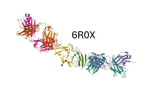
For membrane proteins, FSEC lets us track a GFP-fusion directly in complex mixtures. We use the same selective fluorescence approach to follow labelled protein binders (including antibodies and nanobodies) in mixed samples for both FSEC and Fida studies.
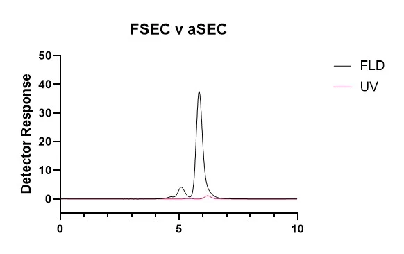
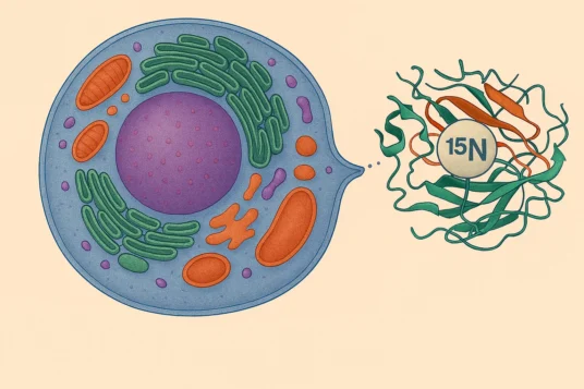
For soluble proteins, the main impact is reduced sample consumption. Switching from standard 280 nm UV detection to intrinsic tryptophan fluorescence achieves a ~40-fold boost in signal.
Recently a client posed an interesting question: could we capture both readouts in a single experiment? They wanted the sensitivity and low sample consumption of intrinsic tryptophan fluorescence, and to unambiguously identify peaks from one labelled component in a complex mixture.
The straightforward workaround was to run each sample twice: once with the FLD tuned to tryptophan, and once to the dye signal (AlexaFluor 488). It would have delivered, but at the cost of doubling both sample consumption and instrument time.
There was one more constraint: our FLD module can’t switch between multiple excitation/emission pairs mid-run. However, if one wavelength is held constant, the other can be scanned – and with a small reduction in scan frequency we can record excitation or emission spectra during the method development phase.
Because many fluorophores share a region of non-selective UV excitation, we ran excitation wavelength scans to map the excitation profile of both fluorophores. This revealed a useful overlap in the low-UV region – one excitation wavelength that could drive both the Tryptophan and AlexaFluor 488 signals at the same time.
We then flipped the experiment and collected emission scans using that low-UV excitation. The result was exactly what we hoped for – characteristic emission spectra for each fluorophore. We could separate intrinsic protein signal from dye-labelled species in a single run.
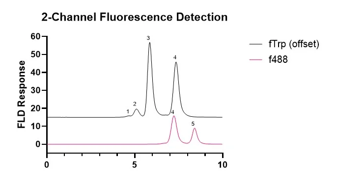
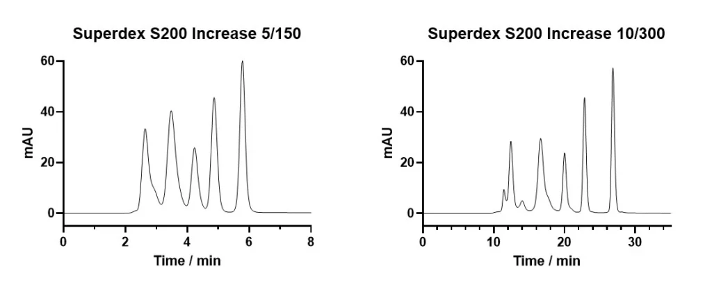
With that in hand, we built a single-run FSEC method that reports both intrinsic and dye-labelled fluorescence. On the example chromatogram below, we could assign peaks 1-3 to unlabelled protein, peak 4 to dye-labelled protein, and peak 5 to unreacted dye – and we could do the peak assignment in real time.
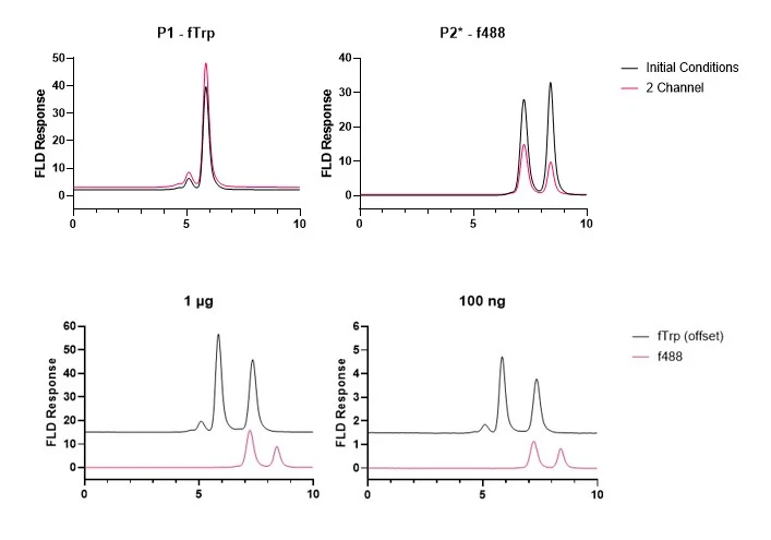
Against the individually optimised settings, the combined method delivered a comparable response on the tryptophan channel, and a slight drop on the AlexaFluor 488 channel. Even so, sensitivity remained excellent: we obtained clean signals from as little as 2 µg/mL labelled protein – an injection of only 10 ng of labelled material.
Although we often use 30 cm SEC columns to maximise resolution, for this rapid method-development work we were grateful to Cytiva for the loan of a Superdex S200 5/150 column. This column can be a great option when high resolution isn’t essential – cutting run time and sample consumption to around a quarter of a standard 10/300 mm column.
Whether you’re interested in applying this approach to your target, or have a complex protein QC problem, contact us and see how our expert Protein Science team can help.


Peak Proteins, NuChem Sciences, and SB Drug Discovery have now fully integrated with Sygnature Discovery.
