

Featured Resources
-
STORM Therapeutics: From Lead to Pre-Candidate Nomination in 18 Months

STORM Therapeutics: From Lead to Pre-Candidate Nomination in 18 Months
Case study
📣From lead to pre-candidate nomination in 18 months. Explore the STORM Therapeutics case study


July 30, 2015

Inobrodib, an exciting, first-in-class oral anti-cancer drug in clinical development by CellCentric, was collaboratively designed, synthesised and supported on its pre-clinical journey by an integrated project team at Sygnature Discovery. Inobrodib is now showing promising results in Phase I and II trials for multiple myeloma and other cancer types.

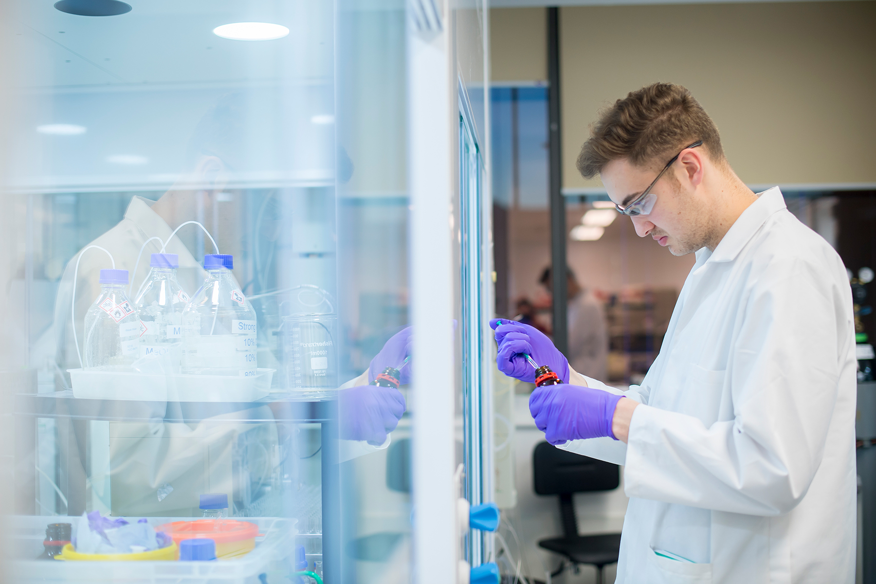
At Sygnature Discovery, we deliver world-leading drug discovery solutions to accelerate your compound from idea to clinic.

Our leadership team brings diverse experience and insight, driving collaboration and innovation across drug discovery.

Explore careers at Sygnature Discovery and join a global team committed to science, collaboration, and integrity.
Inobrodib, an exciting, first-in-class oral anti-cancer drug in clinical development by CellCentric, was collaboratively designed, synthesised and supported on its pre-clinical journey by an integrated project team at Sygnature Discovery. Inobrodib is now showing promising results in Phase I and II trials for multiple myeloma and other cancer types.
At Sygnature Discovery, we deliver world-leading drug discovery solutions to accelerate your compound from idea to clinic.
Our leadership team brings diverse experience and insight, driving collaboration and innovation across drug discovery.
Explore careers at Sygnature Discovery and join a global team committed to science, collaboration, and integrity.
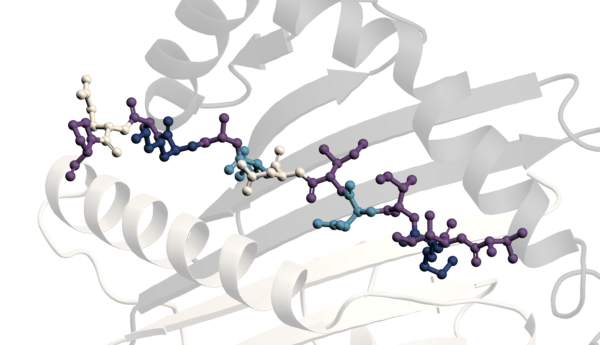
In the absence of Wnt ligands, β-catenin is targeted for proteasomal degradation by a multiprotein “destruction complex” composed of APC (adenomatous polyposis coli), AXIN, GSK-3β (glycogen synthase kinase 3 beta), and CK1 (casein kinase 1). This complex phosphorylates β-catenin at specific serine and threonine residues—primarily encoded by exon 3 of the CTNNB1 gene—marking it for ubiquitination and subsequent degradation (Clevers & Nusse, 2012), preventing its accumulation and nuclear translocation.
Conversely, activation of the Wnt pathway by extracellular Wnt ligands, leads to the destruction complex being inhibited. This inhibition prevents β-catenin phosphorylation and degradation, allowing it to accumulate in the cytoplasm and eventually translocate into the nucleus. Inside the nucleus, β-catenin interacts with TCF/LEF family transcription factors, initiating the transcription of target genes that regulate cell proliferation, survival, and differentiation. This mechanism is essential for normal development and tissue homeostasis but can be hijacked in cancer (Gao, C. et al. 2018).
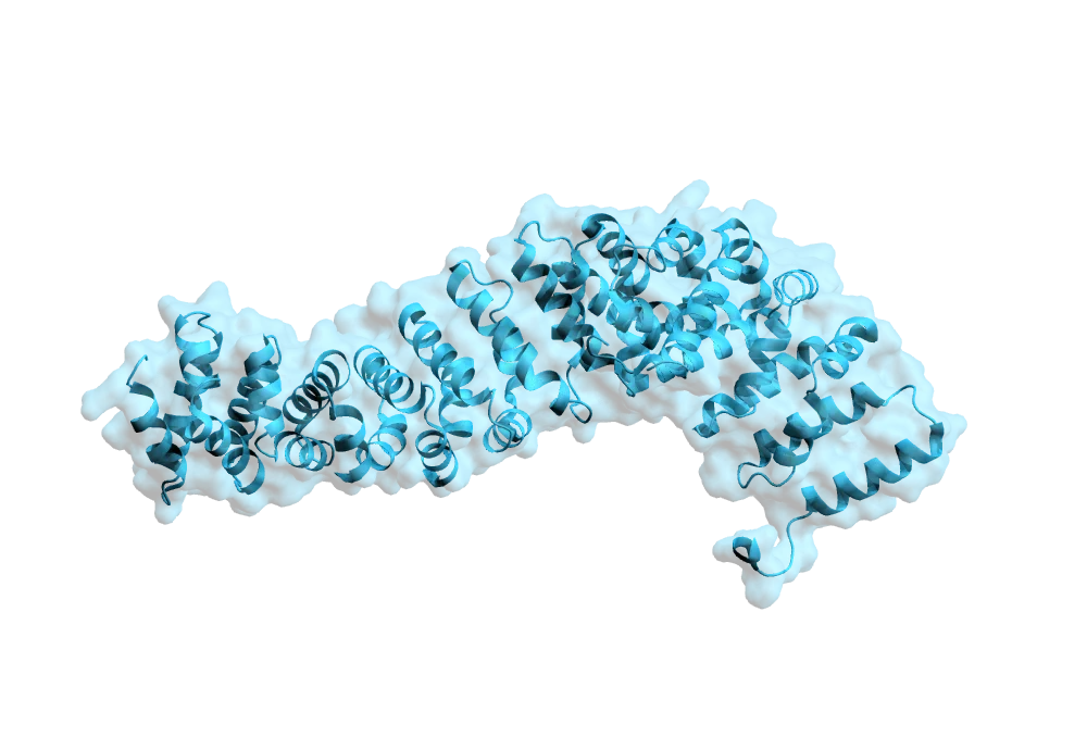
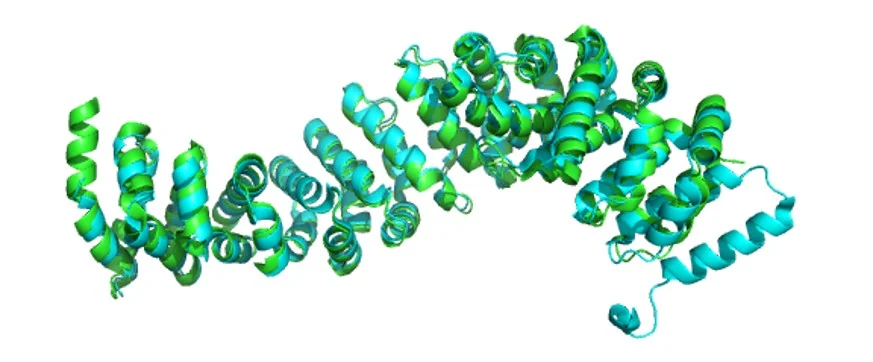
There are multiple CTNNB1 crystal structures deposited within the protein data bank (PDB). Expression, purification, and crystallography of a truncated CTNNB1 construct which included the full C-terminus following the armadillo repeats was achieved by Xing, Y. et al. (2008) (PDB: 2Z6H) (Figure 1. cyan). Whereas, Sampietro, J. et al. (2006) have published the structure of a mutant CTNNB1 crystal structure (PDB: 2GL7) (Figure 1. green). The structures highlight the tight bundles of alpha-helices at the core of the protein providing a scaffold during the Wnt signalling pathway.
Within the Protein Science and Structural Biology department at Sygnature Discovery, we have successfully produced high-quality recombinant CTNNB1 protein to support a wide range of client projects. Our expertise in protein expression and purification enables us to offer multiple construct lengths and tag combinations tailored for diverse downstream applications (Figure 2).
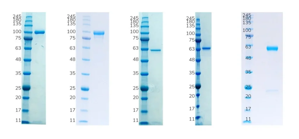
We are looking to build on this success and are currently working to establish a crystallization systems for two of our CTNNB1 constructs. To date we have crystals diffracting to to 3.1 Å but there is a bit more work to be done to generate a robust system….watch this space.
Clever, H. and Nusse, R. Wnt/B-Catenin Signalling and Disease. Cell. 149 (6). 1192-1205. (2012)
Gao, C. et al. Exon 3 mutations of CTNNB1 drive tumorigenesis: a review. Oncotarget 9, 5492 (2018).
Liu, J. et al. Wnt/β-catenin signalling: function, biological mechanisms, and therapeutic opportunities. Signal Transduction and Targeted Therapy 7 (1). 1–23. (2022).
Sampietro, J. et al. Crystal Structure of a beta-Catenin/BCL9/Tcf4 Complex. Molecular Cell. 24 (2). 293 – 300. (2006)
Xing, Y. et al. Crystal Structure of a Full-Length β-Catenin. Structure 16 (3). 478-487. (2008).

Peak Proteins, NuChem Sciences, and SB Drug Discovery have now fully integrated with Sygnature Discovery.
