

Featured Resources
-
STORM Therapeutics: From Lead to Pre-Candidate Nomination in 18 Months

STORM Therapeutics: From Lead to Pre-Candidate Nomination in 18 Months
Case study
📣From lead to pre-candidate nomination in 18 months. Explore the STORM Therapeutics case study


July 30, 2015

Inobrodib, an exciting, first-in-class oral anti-cancer drug in clinical development by CellCentric, was collaboratively designed, synthesised and supported on its pre-clinical journey by an integrated project team at Sygnature Discovery. Inobrodib is now showing promising results in Phase I and II trials for multiple myeloma and other cancer types.

At Sygnature Discovery, we deliver world-leading drug discovery solutions to accelerate your compound from idea to clinic.

Our leadership team brings diverse experience and insight, driving collaboration and innovation across drug discovery.

Explore careers at Sygnature Discovery and join a global team committed to science, collaboration, and integrity.
Inobrodib, an exciting, first-in-class oral anti-cancer drug in clinical development by CellCentric, was collaboratively designed, synthesised and supported on its pre-clinical journey by an integrated project team at Sygnature Discovery. Inobrodib is now showing promising results in Phase I and II trials for multiple myeloma and other cancer types.
At Sygnature Discovery, we deliver world-leading drug discovery solutions to accelerate your compound from idea to clinic.
Our leadership team brings diverse experience and insight, driving collaboration and innovation across drug discovery.
Explore careers at Sygnature Discovery and join a global team committed to science, collaboration, and integrity.
The concept of Targeted Protein Degradation (TPD) covers all techniques that exploit the natural mechanisms of cellular degradation to target a disease-causing protein for destruction.
Targeted protein degradation (TPD) is an emerging therapeutic strategy that hijacks the cell’s ubiquitin–proteasome system to selectively degrade proteins. In this process, ubiquitin tags are attached to lysine residues on target proteins, marking them for destruction. A key player in this cascade is the E3 ligase, which recognizes substrates, polyubiquitinates them, and directs them to the proteasome.
This mechanism can be exploited using bifunctional molecules called proteolysis-targeting chimeras (PROTACs). These molecules simultaneously bind an E3 ligase and a target protein, connected by a linker. Humans have over 600 E3 ligases, but VHL (von Hippel–Lindau) and Cereblon are the most widely used due to their well-characterized binding sites.
At Sygnature Discovery, we have already established production of both VHL and Cereblon which are active in SPR. With VHL-ligand-based PROTACs rapidly advancing—frontrunners now in Phase III trials—the field is full of potential to develop novel therapies across diverse targets. In this blog, we showcase how leveraging our expertise in functional VHL protein production has propelled us to go a step further in TPD.
Within a PROTAC, the binding moieties for the E3 ligase and target protein are critical for substrate recognition, but the linker is equally important. It determines the spatial arrangement and flexibility between the two complexes, ensuring lysine residues remain accessible for polyubiquitination. The linker also influences the PROTAC’s physicochemical and pharmacokinetic properties.
Structural data from ternary complexes (E3 ligase–PROTAC–target) provide valuable mechanistic insights that can guide iterative design for improved efficacy. Similarly, structures of binary complexes (PROTAC bound to either the E3 ligase or target) help optimize binding interactions and linker positioning.
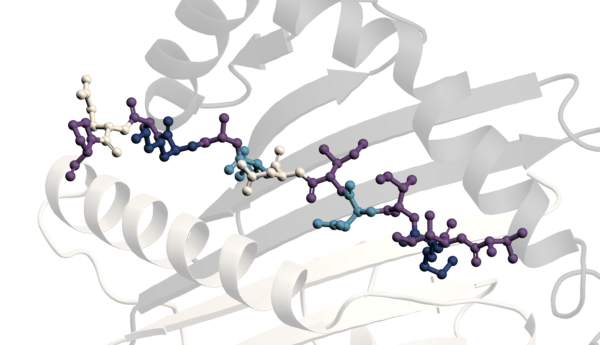
VHL functions within a multi-subunit complex that recruits substrates such as hypoxia-inducible factor (HIF) for ubiquitination and degradation. Together with adaptor proteins Elongin B and Elongin C, VHL forms the VCB complex, a minimal yet powerful tool for TPD development.
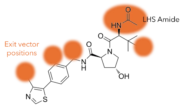
Following the determination of the VCB–HIF structure in 2002, significant efforts focused on designing small molecules that mimic HIF’s binding interface on VHL. Among these, the VH032 scaffold, particularly with a left-hand-side (LHS) amide exit vector, remains the most widely used VHL-binding moiety in degrader development (Figure 1).
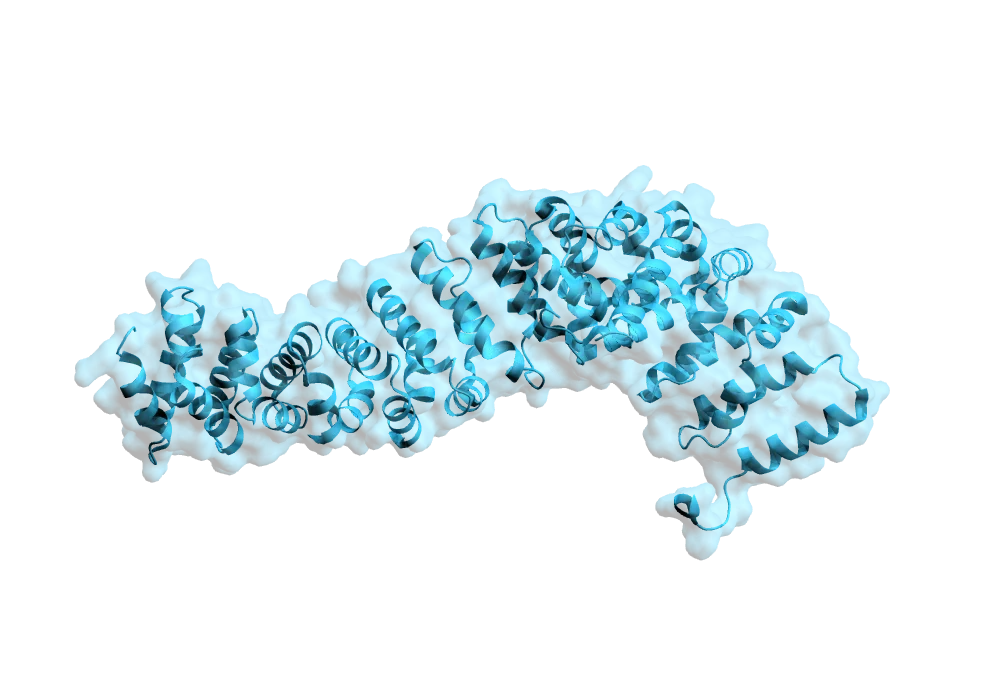
Building upon our expertise in production of highly active, homogenous VCB complex in-house, our team successfully determined the X-ray crystal structure of the VCB complex bound to the tool compound VH032 at 2.3 Å resolution (Figure 2a). The heterotrimeric VCB complex crystallized under conditions optimized from literature and broad screening. The resulting crystals contained two heterotrimers per asymmetric unit, each with VH032 bound to VHL.
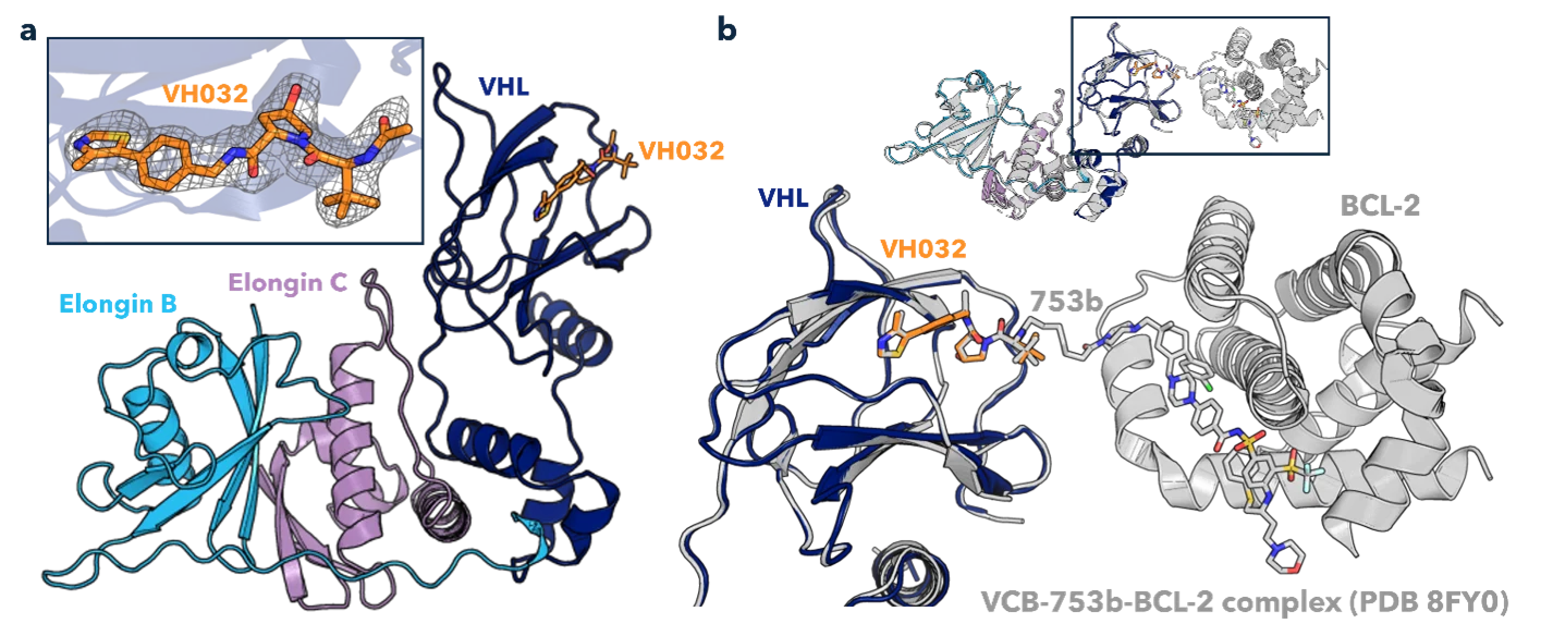
A comparison of our in-house VCB-VH032 structure with a published crystal structure of VCB bound to a VH032-based PROTAC in complex with BCL-2 shows excellent superimposition (Figure 2b). This not only confirms the integrity of our VCB complex but also allows us to test the DMSO tolerance of crystals- opening avenues for soaking or co-crystallization with additional ligands.
If you would like to learn more about our protein services or other services, please get in touch.

Peak Proteins, NuChem Sciences, and SB Drug Discovery have now fully integrated with Sygnature Discovery.
