

Featured Resources
-
STORM Therapeutics: From Lead to Pre-Candidate Nomination in 18 Months

STORM Therapeutics: From Lead to Pre-Candidate Nomination in 18 Months
Case study
📣From lead to pre-candidate nomination in 18 months. Explore the STORM Therapeutics case study


July 30, 2015

Inobrodib, an exciting, first-in-class oral anti-cancer drug in clinical development by CellCentric, was collaboratively designed, synthesised and supported on its pre-clinical journey by an integrated project team at Sygnature Discovery. Inobrodib is now showing promising results in Phase I and II trials for multiple myeloma and other cancer types.

At Sygnature Discovery, we deliver world-leading drug discovery solutions to accelerate your compound from idea to clinic.

Our leadership team brings diverse experience and insight, driving collaboration and innovation across drug discovery.

Explore careers at Sygnature Discovery and join a global team committed to science, collaboration, and integrity.
Inobrodib, an exciting, first-in-class oral anti-cancer drug in clinical development by CellCentric, was collaboratively designed, synthesised and supported on its pre-clinical journey by an integrated project team at Sygnature Discovery. Inobrodib is now showing promising results in Phase I and II trials for multiple myeloma and other cancer types.
At Sygnature Discovery, we deliver world-leading drug discovery solutions to accelerate your compound from idea to clinic.
Our leadership team brings diverse experience and insight, driving collaboration and innovation across drug discovery.
Explore careers at Sygnature Discovery and join a global team committed to science, collaboration, and integrity.
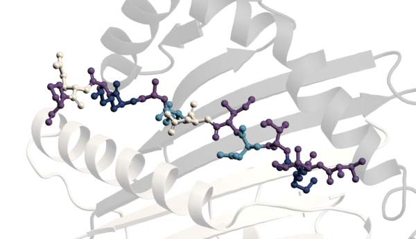
CLC-Ka and CLC-Kb are members of the CLC family of chloride channels and transporters, crucial for maintaining chloride ion homeostasis across cell membranes. CLC-Ka is predominantly found in the thin ascending limb of Henle’s loop, while CLC-Kb is expressed in both the thick ascending limb of Henle’s loop and the distal convoluted tubule. These channels facilitate the reabsorption of chloride ions from the filtrate back into the bloodstream, essential for maintaining the body’s fluid and electrolyte balance. Additionally, these channels are expressed in the inner ear, where they are vital for normal hearing and balance.

Given their vital role in renal function, CLC-Ka and CLC-Kb present promising targets for therapeutic intervention in various kidney-related disorders. Modulation of these channels can potentially treat conditions characterized by improper chloride handling and disrupted electrolyte balance, such as hypertension, edema, congestive heart failure, Bartter syndrome, and chronic kidney disease.
Our specialized electrophysiology screening services offer cutting-edge solutions for drug discovery targeting CLC-Ka and CLC-Kb channels. Utilizing state-of-the-art technology, we provide comprehensive and high-throughput assays to evaluate the efficacy of potential drug candidates.
HEK cells stably expressing CLC-Ka and CLC-Kb were produced by Sygnature Discovery. Whole cell patch-clamp experiments were carried out at room temperature using multi-hole chips on the SynchroPatch 384i automated electrophysiology platform. Currents were elicited by using repeated ramps, steadily increasing from -140 mV to + 60 mV over 500 ms, from a holding potential of – 30 mV.
Data analysis was performed using Data Control 384 V2.3 (Nanion) and GraphPad Prism V10.1.
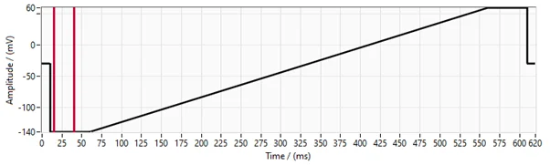
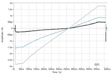
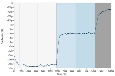
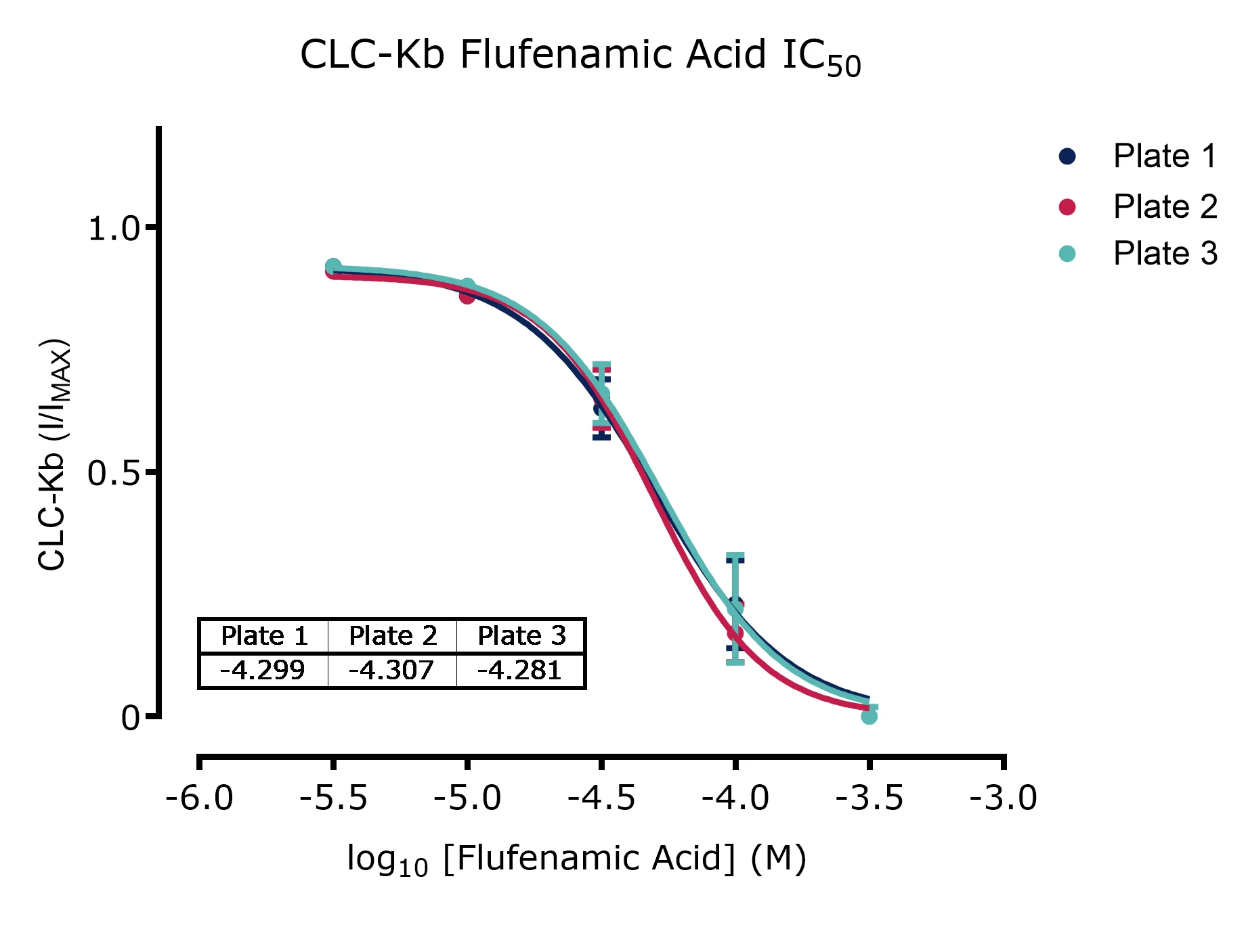
Figure 4. Representative current trace, time course and concentration response curve showing the effect of inhibitor against CLC-Kb and reproducibility across 3 plates. An example of superimosed CLC-Kb current traces under control conditions (grey line), in the presence of 30 μM Flufenamic acid (blue line) and a saturating concentration of 300 μM Flufenamic acid (black line).


Peak Proteins, NuChem Sciences, and SB Drug Discovery have now fully integrated with Sygnature Discovery.
